

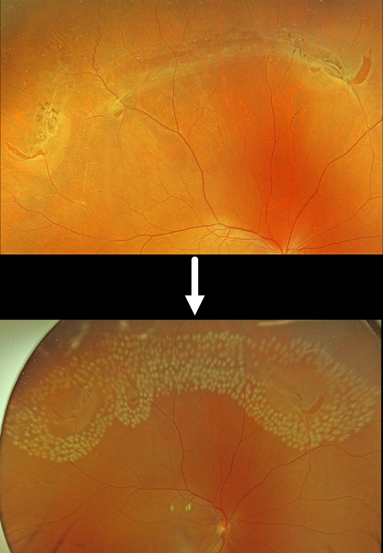
This 55YO male was referred for an acute posterior vitreous detachment with symptomatic retinal tears. Vision was 20/25.
Optos color RGB imaging shows several patches of lattice degeneration extending from the 10 to 1:30 o’clock meridians associated with multiple retinal tears. Some inferior vitreous hemorrhage was noted (not shown). Immediate prophylactic laser photocoagulation was applied to all surrounding the lattice and breaks.
Lattice lesions have a pocket of liquefied vitreous overlying thinned retina with a firm area of surrounding vitreoretinal adhesion. White, fibrosed vessels often crisscross the lesions, giving them their name, “lattice.” This firm vitreoretinal adhesion, along with the often-present atrophic holes, is what can predispose these eyes to retinal breaks and detachment. Although prophylactic treatment is rarely needed, symptomatic breaks, as in our patient, need prompt retinopexy.



