

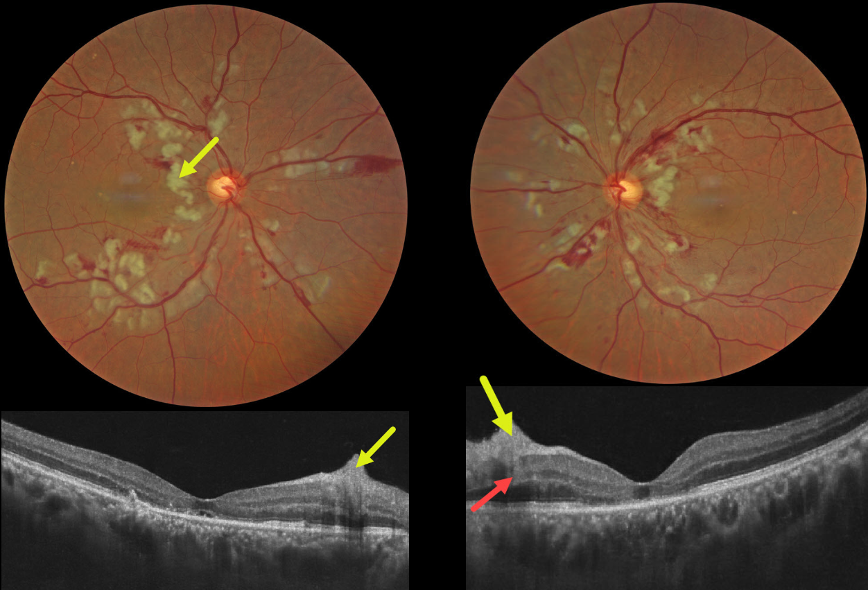
This 55YO male presented with sudden bilateral vision loss that developed immediately following a motor vehicle accident, which included blunt thoracoabdominal trauma and transient loss of consciousness. Vision was 20/200 OU.
Color photography shows bilateral inner and mid-retinal patches of white ischemic retina. The nerve fiber layer lesions appear as inner retinal hyperreflective thickening on OCT. Mid-retinal hyperreflectivity involving the outer plexiform/inner nuclear/outer plexiform layers is noted nasally OS. The outer photoreceptor bands are variably disorganized OU, with a small amount of subfoveal subretinal fluid OD and a foveal cyst OS.
Learning Points:
Purtscher retinopathy was first described by Otmar Purtscher in 1910. Although originally described in a man who fell from a tree with cranial trauma, these white patches of retinal ischemia and hemorrhages are found in numerous other etiologies, including pancreatitis, chest trauma, and collagen vascular diseases. The findings are called Purtscher retinopathy when due to trauma, and Purtscher-like retinopathy when seen from other causes.
The underlying cause of Purtscher is unknown, but it is thought to represent leukoembolization by complement activation. The areas of ischemia include more superficial nerve fiber layer infarcts (cotton wool spots) and deeper ischemia involving the middle retinal layers. This recently described retinal ischemic phenotype, paracentral middle maculopathy (PAMM), occurs as an idiopathic entity or in other ocular disorders, including Purtscher retinopathy, diabetic retinopathy, retinal artery and retinal vein occlusions, and acute macular neuroretinopathy (see Retina 2015;35:1921-1930).



