

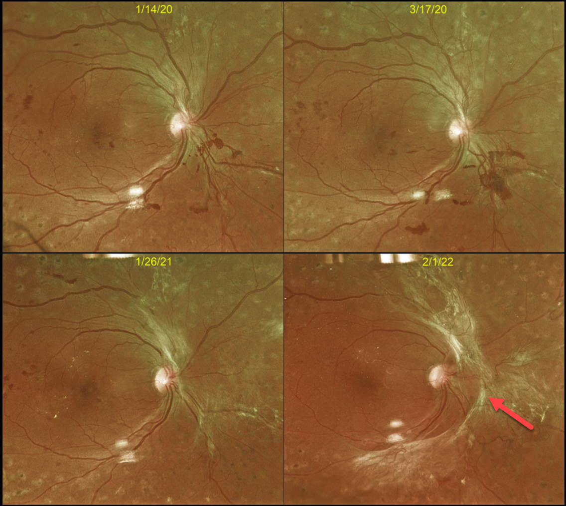
This 43YO male with type 2 diabetes initially responded beautifully in 2016 to full panretinal photocoagulation (PRP) for proliferative diabetic retinopathy (PDR). However, he presented on 3/17/20 with some new preretinal blood (Optos color RG image), and fill-in PRP was applied.
Over the course of 2 years, progressive fibrotic neovascularization with extramacular traction developed. Despite the traction, vision remained at 20/40, and careful observation was recommended. We were hesitant to add further laser or anti-VEGF therapy for fear of causing increased traction and ‘crunch’ (Tan et al, Surv Ophthalmology 2021;66:926-932). When last examined on 4/3/24, the retinal findings remained unchanged, and vision was 20/30 (not shown).
Although PRP is often a ‘one and done’ procedure, this is not always the case (Gonzalez et al, Ophthalmology 2021;128:1448-1457). Our patient is a perfect example of why these patients require lifelong monitoring even if their disease appears quiescent.



