

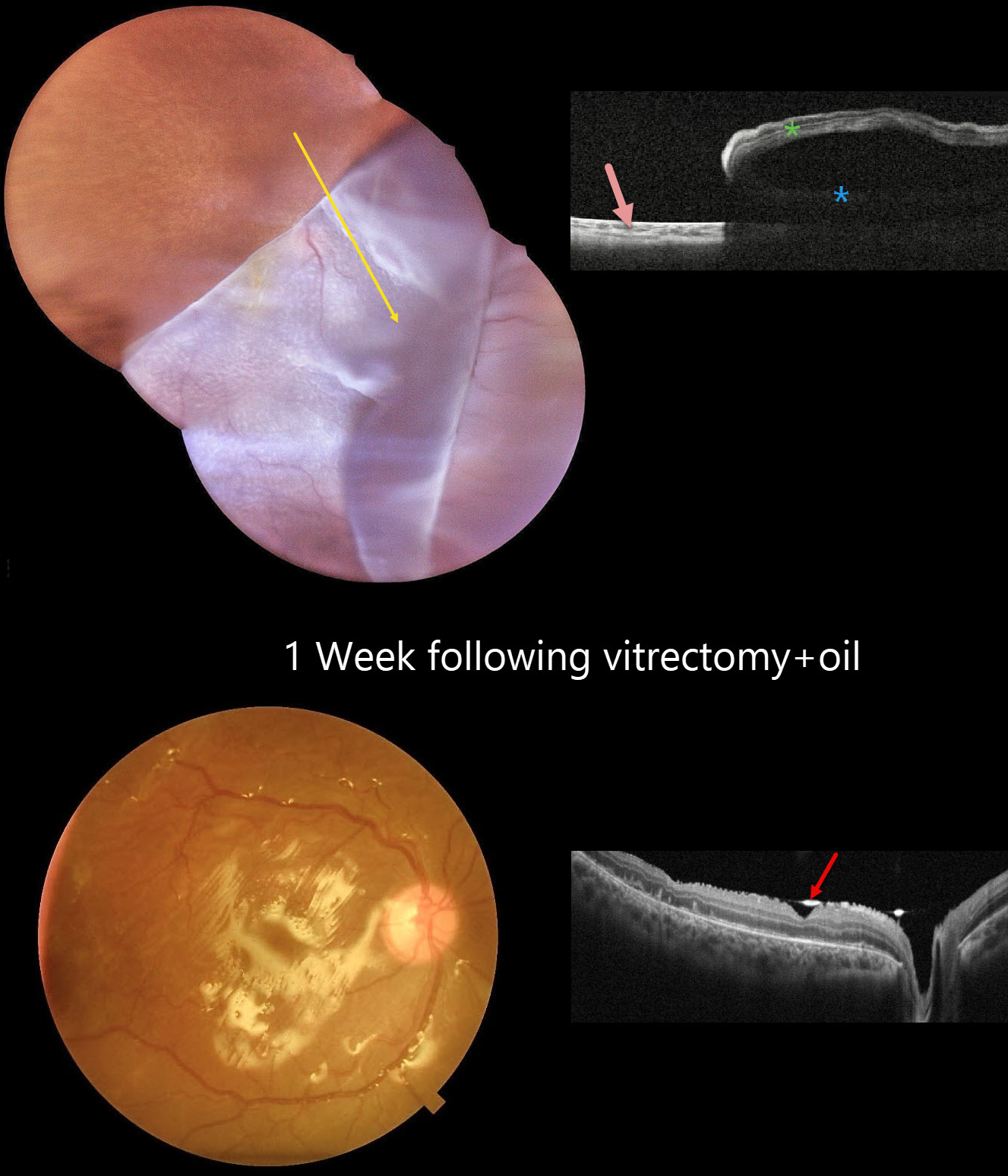
This 12YO boy presented with 3 weeks of vision loss in his right eye immediately following blunt trauma. Vision was hand motion OD and 20/20 in his normal OS.
Color photography shows a superotemporal giant retinal tear that has folded over the optic nerve and macula inferonasally. The yellow macular xanthophyll is visible just inferotemporal to the nerve. OCT scanning shows bare RPE superotemporally, the underlying detachment, and the overlying inverted retina.
Emergent surgical repair with pars plana vitrectomy was scheduled. One week postoperatively, the retina is completely attached. The hyperreflective oil-retinal interface is visible overlying the fovea and optic nerve.



