

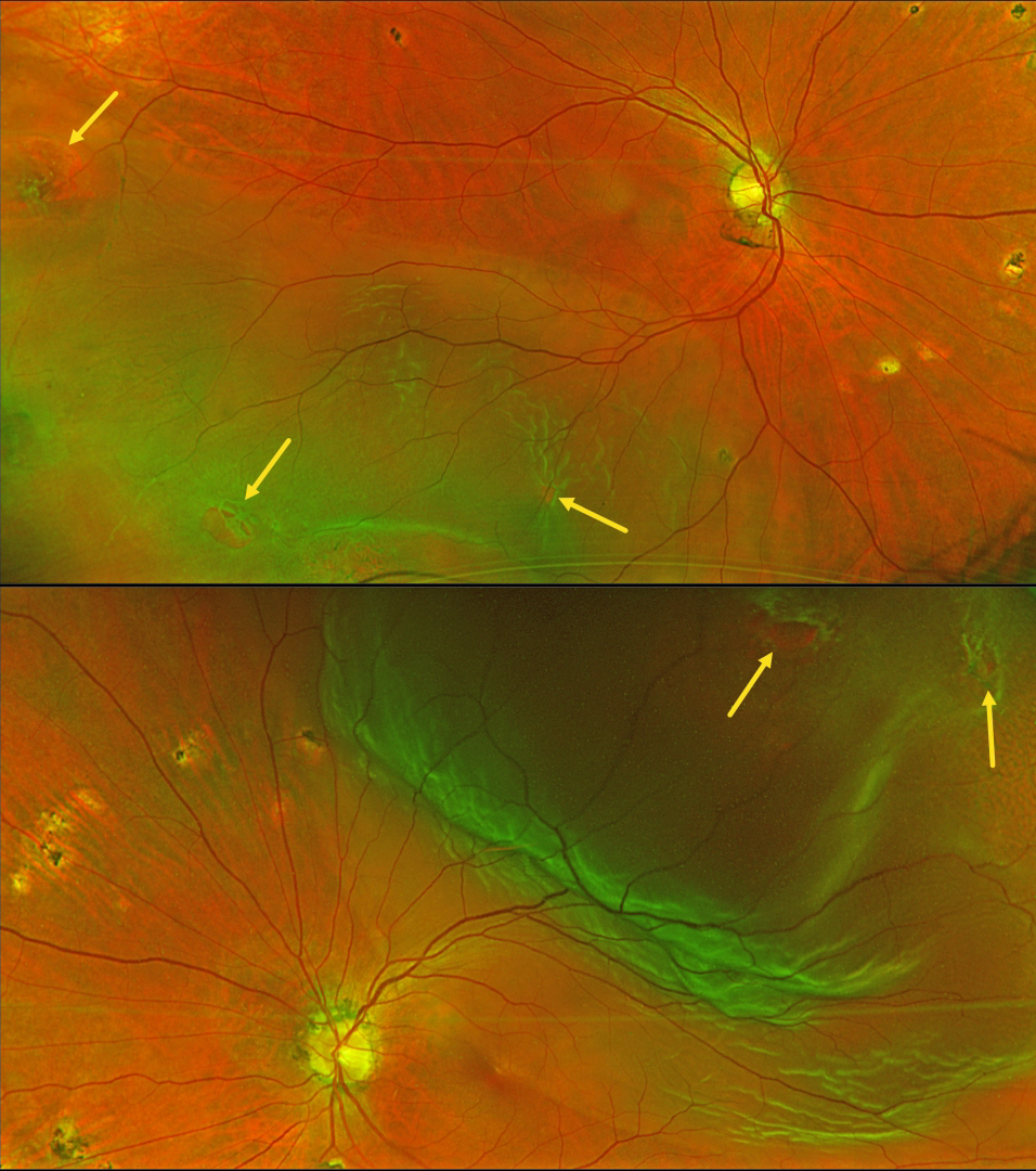
This 60YO male presented with 2 weeks of floaters in his left eye. Vision was 20/25 OD and 20/30 OS. Optos color RG imaging shows bilateral rhegmatogenous retinal detachments (RRD) with numerous breaks. The asymptomatic right detachment extended into the inferotemporal macula and split the left fovea. Scattered ocular histoplasmosis scars are present bilaterally.
Learning Points:
Patients with a RRD have a lifelong 10% risk of detachment in their fellow eye. Only a very small percentage will present with simultaneous detachments (Singh et al, Retina 2019;39:1504-1509).
Although the surgical success for each eye should be no different from that in patients presenting with unilateral detachments, treating the patient (not the eye) is more complicated. If one chooses pars plana vitrectomy (PPV), the postoperative gas bubble can cause severe, temporary vision loss. If both eyes receive PPV close together in time, the patient will be functionally blind for several weeks to months. We therefore find it helpful in these cases to treat the first eye with either silicone oil (with postoperative glasses) or perfluoro-octane liquid (followed by surgical removal a few weeks later). We can then repair the second eye within a few weeks of the initial eye. If possible, scleral buckling is also a good choice since no gas bubble is needed.
Emergent pars plana vitrectomy surgery was scheduled for our patient’s left detachment. We will follow the right eye closely perioperatively and will be forced to operate sooner if the detachment spreads towards the macular center. Otherwise, the right detachment will be repaired once the intraocular bubble is less than 50% in his left eye.



