

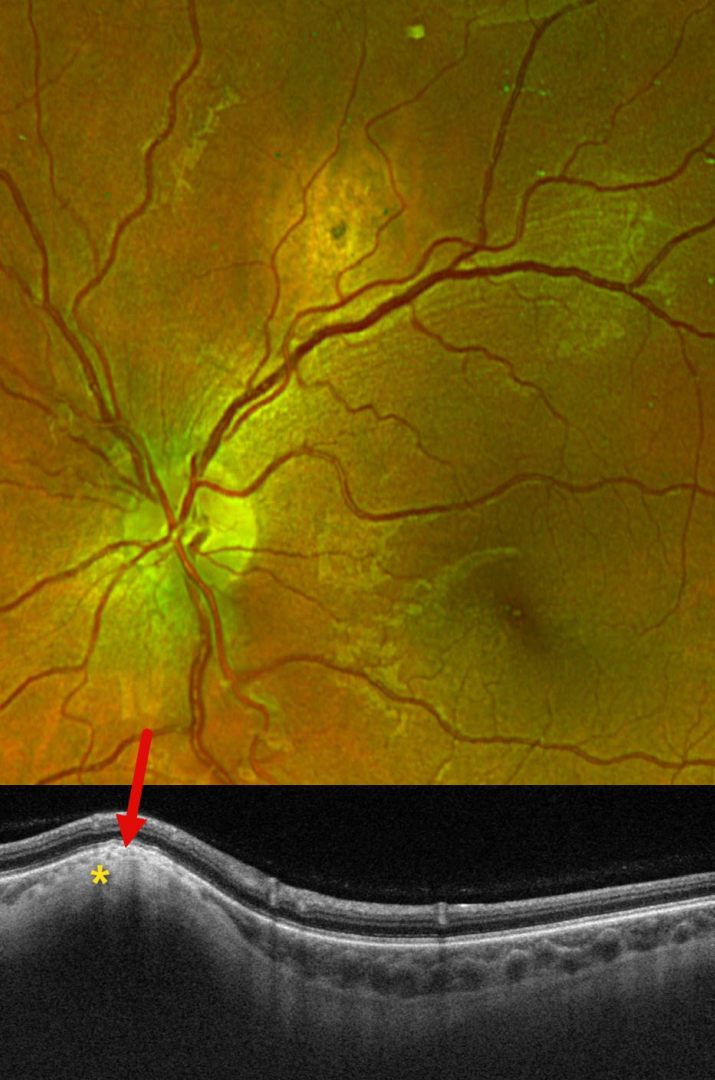
This 29YO female presented with this asymptomatic amelanotic lesion above the right superotemporal arcade. Optos color RG imaging shows an apparent mostly depigmented chorioretinal scar. However, Triton swept-source OCT scanning shows hyperreflective scleral thickening, which compresses the overlying choroid and elevates the retina.
Learning Points:
Focal scleral nodule, previously known as solitary idiopathic choroiditis (SIC) and unifocal helioid choroiditis, presents as a single, yellow-white, elevated nodular, avascular subretinal lesion. On OCT, it arises from the inner sclera, causing marked choroidal compression. These lesions require no treatment, although they may rarely show slight growth (Fung and Li, Retinal Cases & Brief Reports 2024;18:526-528).



