

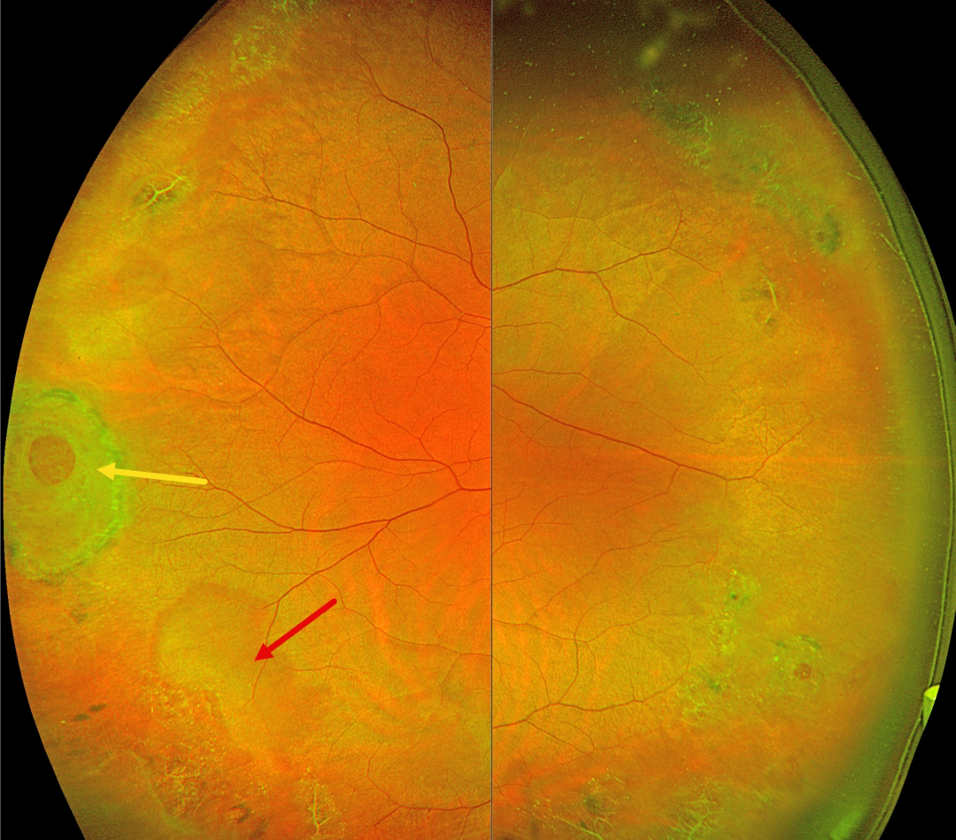
This 42YO female was referred for asymptomatic peripheral retinal findings. Vision was 20/20 OU.
Optos color RG imaging shows extensive bilateral temporal lattice lesions with atrophic holes. A larger hole with surrounding pigment is seen in the right eye, along with a nearby area of white without pressure.
Learning Points:
Lattice lesions have a pocket of vitreous liquefaction overlying the thinned retina with a firm vitreo-retinal adhesion along their borders. White, fibrosed vessels often crisscross the lesions, which leads to the name, lattice. This firm vitreoretinal adhesion, along with the often-present atrophic holes, is what can predispose these eyes to rhegmatogenous retinal detachment (RRD).
Retinal detachment occurs in 1 in 10,000 people per year. Lattice is found in 10% of the general population, especially in high myopes. Since about one-third of patients with retinal detachment have lattice as the cause, the question always arises: Should prophylactic retinopexy be performed? The short answer is that asymptomatic lattice should generally not be treated.
Byer, in a Long-term natural history study of 423 untreated eyes, concluded that prophylactic treatment with or without holes in phakic non-fellow eyes should not be performed (Ophthalmology 1989;96:1396-1402). Folk et al (Ophthalmology 1989;96:72-79, Retina 1990;10:165-169) found no compelling evidence for prophylactic treatment of phakic fellow higher-risk eyes with more extensive lattice or higher myopia. They also found that some eyes developed subsequent non-lattice breaks outside the prophylactic treatment. A recent Cochrane systematic review by Wilkinson concluded that asymptomatic breaks and lattice should not be treated (Cochrane Database of Systematic Reviews 2014;9;CD003170) and the most recent 2019 American Academy of Ophthalmology Preferred Practice Pattern also cautions against prophylactic treatment.
A recent study by Curran et al recommended prophylactic treatment of lattice in fellow retinal detachment eyes, but this was in a retrospective, uncontrolled case series (Retina 2024;44:63-70). Another recent report by Kazan et al (Journal of VitreoRetinal Diseases 2024;8:381-387), in an uncontrolled retrospective series of eyes with “high-risk” lattice, found few post-laser detachments and concluded that treatment “may be effective in decreasing progression to RD…” But 3% of their treated eyes developed RRD over a mean 3- to 4-year follow-up period, which is higher than expected from the natural history of untreated lattice. So, our interpretation of their results is that prophylactic treatment may have actually increased the risk for detachment.
Stickler syndrome is a usually autosomal-dominant genetic disorder characterized by defective vitreous collagen (types II, IX, and XI). It is the most common cause for inherited rhegmatogenous retinal detachment and is the exception to the rule against prophylactic treatment. Prophylactic encircling laser extending from the pre-equator to the ora should be strongly considered since it lowers the risk for retinal detachment and giant retinal tears (Linton et al, Retina 2023;43:88-93).



