

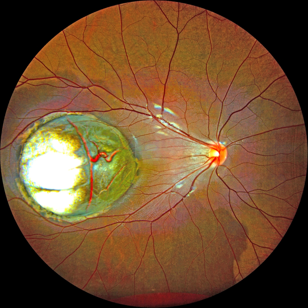
This healthy 5YO boy’s parents noticed that his right eye was wandering for a few weeks. Vision was 20/200 OD and 20/20 in the normal OS.
Color fundus photography shows a large choroidal coloboma occupying the central and temporal macula. The underlying outer choroidal vessels are visible. His systemic examination was normal. Serologic testing for toxoplasmosis and cytomegalovirus was negative.
Learning Points:
Choroidal colobomas are most commonly due to failure of the optic vesicle and choroidal fissure to close during embryonic development and are usually found inferonasally. They can present as solitary or multiple lesions. The differential diagnosis for congenital macular coloboma also includes post-inflammatory lesions (toxoplasmosis, cytomegalovirus, and Zika) and retinal dystrophies, including North Carolina macular dystrophy. The retina within the coloboma is variably thinned, increasing the risk of rhegmatogenous retinal detachment (RRD).



