

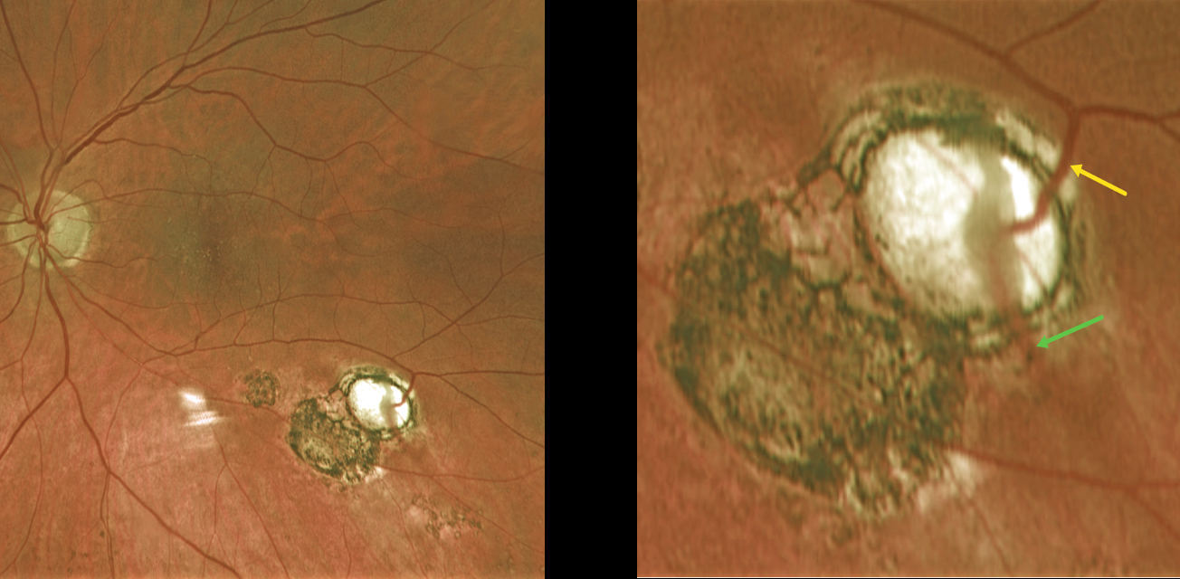
This 60YO female was referred for asymptomatic scarring in her left eye. Vision was 20/50 OD and 20/40 OS due to early bilateral cataracts. Small macular drusen were noted bilaterally.
Optos color RG imaging of her left shows multifocal variably pigmented scars below the inferotemporal arcade. A magnified inferotemporal view shows a retinal vein diving into an atrophic scar and directly connecting to a choroidal vein.
Learning Points:
Under normal conditions the retinal and choroidal circulations remain separate and distinct. Rarely, the two circulations directly connect with each other through a chorioretinal anastomosis. These can occur in chorioretinal scars (typically from inactive toxoplasmosis lesions as in this patient), in macular telangiectasia, and in disciform scars. We felt our patient’s findings most consistent with inactive toxoplasmosis scarring.



