

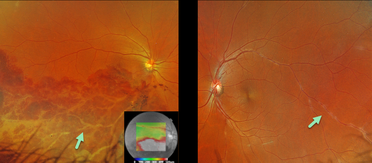
This healthy 36YO male presented with 1 week of unilateral vision loss. Vision was 20/400 OD and 20/20 OS.
Optos color RGB imaging OD shows a major inferotemporal branch retinal vein occlusion (BRVO) with severe confluent retinal hemorrhages and a frosted appearance to the major vessels. The occlusion does not seem to arise from a more proximal arteriovenous (AV) crossing. A small area of peripheral superotemporal periphlebitis is noted OD, with more extensive periphlebitis OS. Triton swept-source OCT shows inferior macular fluid.
BRVOs nearly always arise at an AV crossing, where a thickened arterial vessel compresses the underlying vein. Sarcoidosis is the classic exception where a BRVO is not caused by an AV crossing, but by an inflammatory narrowing of the involved vein. In addition, our patients’ segmental periphlebitis (‘candle wax drippings’) is virtually pathognomonic for sarcoidosis as well.
A sarcoidosis workup was recommended, although the patient failed to show for his testing and was immediately lost to follow-up.



