

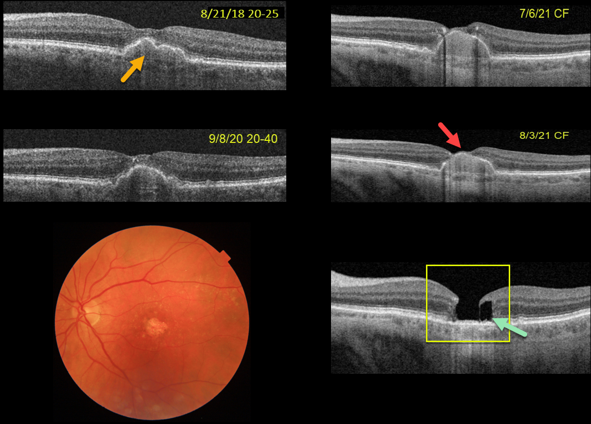
This 75YOF was seen in 2018 with non-neovascular AMD and a central pigment epithelial detachment (PED). Vision was 20/25. She returned on 9/8/20 with 20/40 vision with mild enlargement of the PED.
She complained of decreased vision when seen nearly one year later. Vision had dropped to 20/80. The PED had enlarged to involve full-thickness retina. She elected for an Eylea injection in hopes of possibly treating an occult macular neovascularization vs an avascular PED.
On 7/6/21, vision decreased to counting fingers although the OCT looked fairly stable. An additional Eylea injection followed by half-fluence PDT was given. On 8/3/21, the dome of the PED now extended through the inner retina and vision remained at counting fingers.
On 1/4/22, the PED had completely collapsed, with secondary retinal atrophy and a degenerative lamellar macular hole (LMH).
The progression of her PED through full-thickness retina and subsequent collapse with secondary retinal/RPE atrophy and a LMH are all quite unusual. Fortunately her right eye is 20/20 with intermediate dry AMD.



