

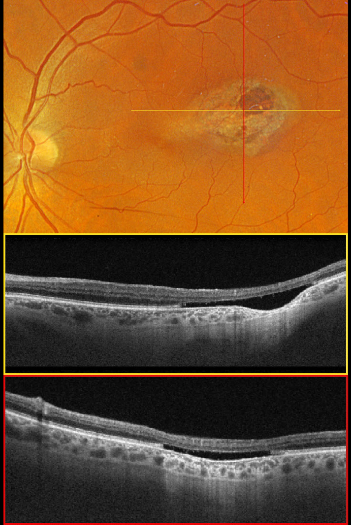
This 66YO male was referred for this unilateral, asymptomatic, variably pigmented lesion in his right eye. Vision was 20/30.
Optos color RGB imaging shows a torpedo-shaped lesion in the temporal macula. Triton swept-source OCT shows an outer retinal cavitation with loss of retinal tissue deep to the external limiting membrane. There is also focal choroidal thinning within the temporal aspect of the lesion.
Learning Points:
Torpedo maculopathy was originally described by Gass as a solitary hypopigmented nevus of the RPE (Arch Ophthalmology 1992;1358-1359). These benign, unilateral lesions are teardrop-shaped and almost always located along the horizontal meridian of the temporal macula. OCT scanning shows attenuation of the RPE and outer retinal layers. Wong et al classified these lesions into type 1 (‘mild outer retinal disturbance’) and type 2 with an outer retinal cavitation as seen in our patient (Clinical Exper Ophthalmology 2015;43:342-348). Vision is almost always normal, although there is a small risk for macular neovascularization. There are no known systemic associations.



