

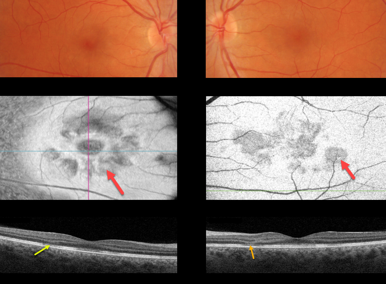
This 41YO female presented with acute bilateral central vision loss. Fundus photography shows very faint reddish-brown wedge-shaped lesions radiating from each fovea.
Swept-source en face imaging of the outer retina shows multiple hyporeflective wedge-shaped lesions radiating from the macular centers.
OCT B-scanning shows bilateral areas of outer nuclear layer thinning with hyperreflectivity associated with disruption of the ellipsoid zone and outer segment layers.
Learning Points:
This case highlights many characteristic features of acute macular neuroretinopathy (AMN), an idiopathic disorder likely caused by compromise of the deep retinal capillary plexus.
Multicolor and infrared imaging often highlight the typical wedge-shaped AMN lesions more effectively than color fundus photos, although the en face OCT images in our patient beautifully capture these pathognomonic findings.
Most patients are white, non-Latino females with a median age of 30 years. The most common associations are a flu-like prodrome and oral contraceptive use.
Presenting acuity is usually good, coinciding with sparing of the sub-foveal areas. Roughly half of AMN cases are bilateral. There is typically moderate visual improvement over several months without treatment.



