

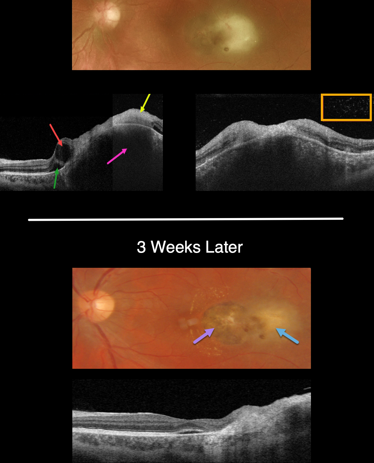
This 34YO male presented with recent vision loss of counting fingers vision in his left eye. He gave a history of prior ocular toxoplasmosis.
Optos color imaging shows opaque and white retina temporal to the fovea. Swept-source OCT shows the clinical retinitis involving full-thickness retina and is markedly hyperreflective. The underlying choroid is markedly thickened. The temporal fovea has a large cyst with a small amount of subretinal fluid. Overlying vitreous cells are noted.
The patient was started on a six-week course of oral Bactrim-DS BID. Laboratory testing for toxoplasmosis showed high IgG and negative IgM titers. ACE, lysozyme and QuantiFERON TB were negative.
The chorioretinal inflammation improved at each visit. When last seen 3 weeks into therapy, the likely pre-existing temporal pigmented toxoplasmosis scar is seen with resolving temporal retinitis. A few dots of retinal blood are seen, and faint lipid is noted nasally.
OCT scanning shows resolved vitreous cells and markedly decreased choroidal thickening.
Learning Points:
Toxoplasmosis, the most common cause of infectious chorioretinitis in humans, is caused by the protozoan parasite, Toxoplasma gondii.
It is most commonly transmitted through infected felines or by eating raw meat. The majority of infections are acquired, although it can also be transmitted congenitally.
The body’s immune system is unable to completely kill the organism, which often lies dormant within a chorioretinal scar for years. When the immune balance favors the encysted organism, active chorioretinitis develops. This usually resolves spontaneously within 6 weeks.
If the optic nerve or macular are threatened, a six-week course of Bactrim DS is recommended (see Ophthalmology 2004;112:1876-1882). Oral prednisone can be added as well to help with severe inflammation.



