

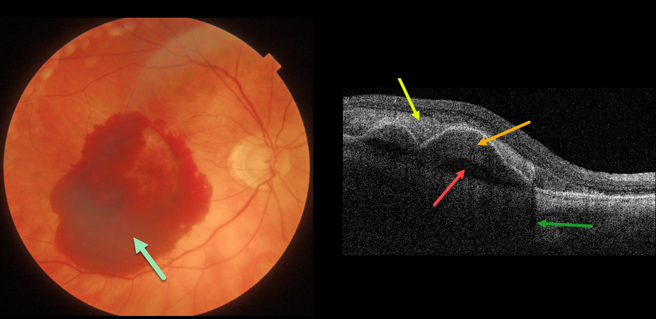
This 84 YO female with macular degeneration presented with acute vision loss of counting fingers vision in her right eye from neovascular AMD (nAMD).
Fundus photography shows a large central subretinal hemorrhage. Swept-source OCT shows the hyperreflective subretinal blood, which shadows the underlying choroid. Sub-RPE blood vs a type-1 (located beneath the RPE) macular neovascularization (MNV) overlies a prechoroidal cleft.
Anti-VEGF therapy was begun.
Learning Points:
A prechoroidal cleft is a hyporeflective space between the RPE and Bruch’s membrane. These are seen in up to about 20% of treated eyes with nAMD, particularly with type 3 neovascularization (intraretinal MNV) and polypoidal choroidal vasculopathy.
They are usually associated with worse visual acuity due to potential complications including RPE tear and subretinal hemorrhages (Kim et al, Retina 2017;37:2047-2055).



