

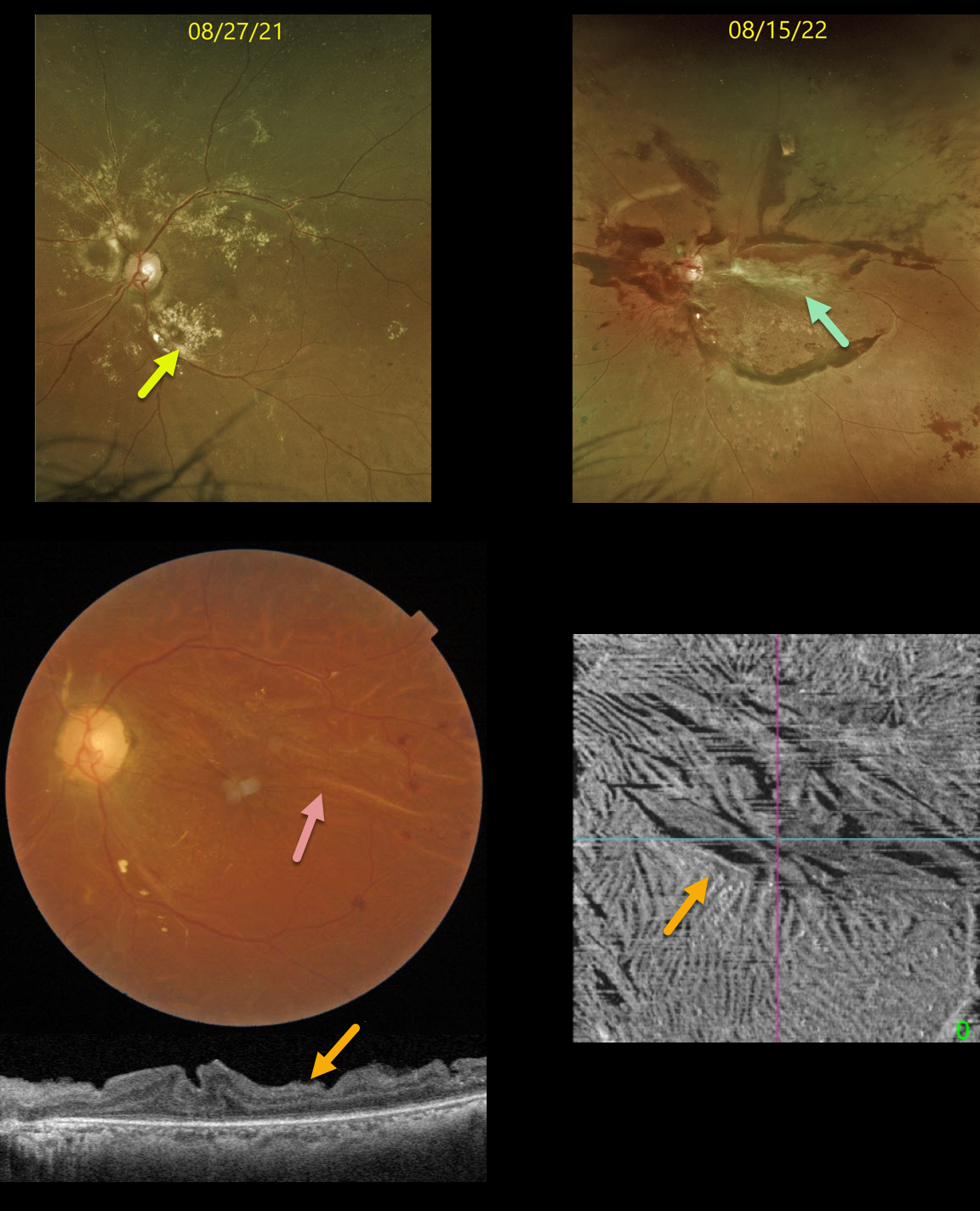
This 38YO diabetic female was initially seen on 8/27/21 with extensive dry macular lipid in her left eye and proliferative diabetic retinopathy. Vision was 20/40. Panretinal photocoagulation was recommended.
Unfortunately, she was lost to follow-up until she returned on 8/15/22 with 20/200 vision from a traction retinal detachment. The lipid spontaneously absorbed.
Vitrectomy surgery was performed, and three weeks later, the retina was completely attached with 20/70 vision.
Color imaging of the macula shows variable retinal folds centrally and superiorly.
OCT B-scan and en face of the inner retina variably show the dramatic inner retinal folds caused by the prior traction. We expect these to improve over time, but will probably never be completely resolved.
Learning Points:
Inner retinal folds, although usually less dramatic than in our patient, are fairly common following vitrectomy for diabetic TRD. They often fail to completely resolve, and vision can be quite good despite the OCT findings.



