

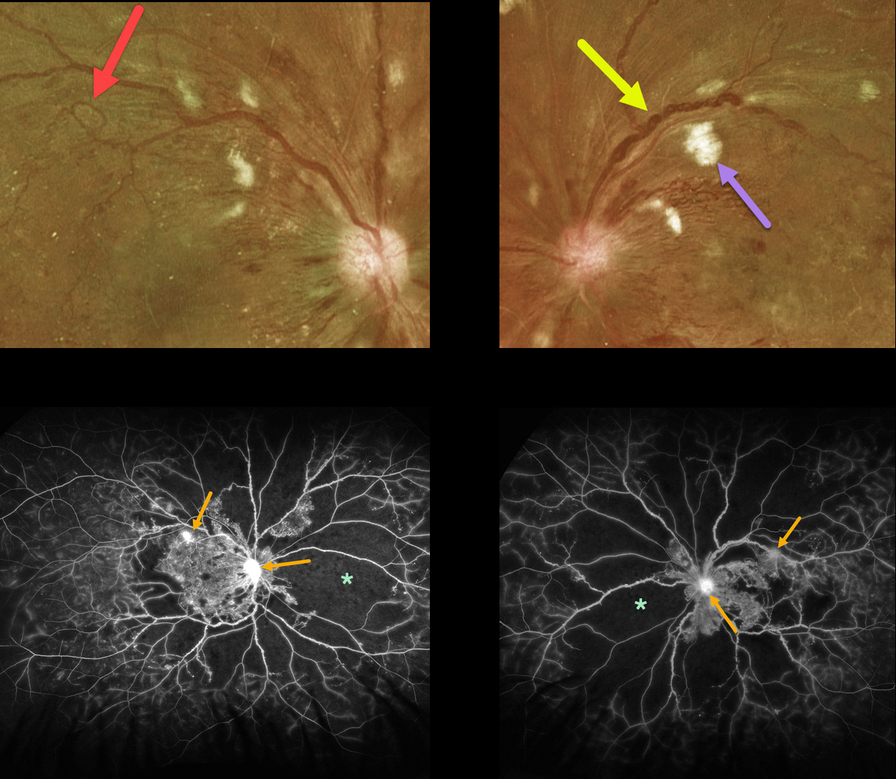
This 33YO diabetic male presented with vision of 20/30 OD and 20/100 OS.
Optos color imaging shows bilateral severe posterior pole retinal hemorrhages and nerve fiber layer infarcts. Florid disc neovascularization is noted bilaterally. Marked venous beading is noted in each eye, and a large venous loop is noted in his right eye. The peripheral retina is also markedly ischemic.
Fluorescein angiography shows marked severe ischemia. The venous beading is much more global angiographically, with extensive venous staining of the irregularly beaded veins. Despite the ischemia, just a few areas of neovascularization are noted in each eye.
Vision is good OD since the foveal avascular zone is relatively normal, and vision is poor OS due to foveal ischemia.
Panretinal photocoagulation was recommended for each eye.



