

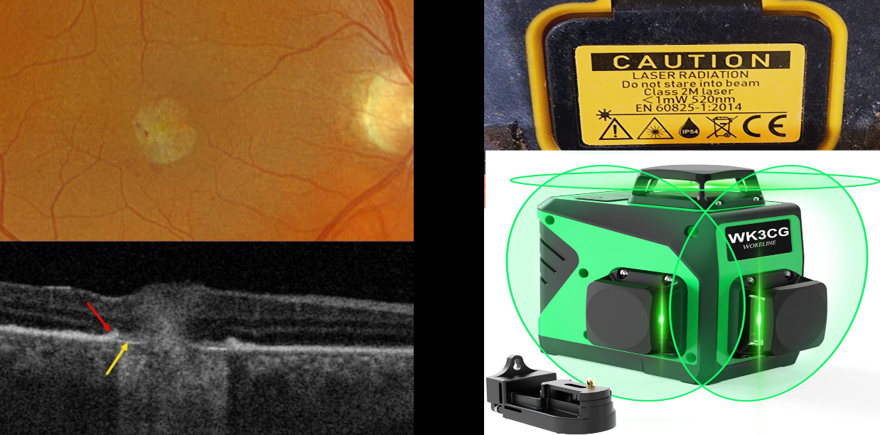
This 46YO male presented with a several-week history of unilateral vision loss. Vision was 20/20 in his normal left eye and counting fingers in his right eye.
Optos color RGB imaging shows a mostly round foveal scar with a few temporal blood dots. Triton swept-source OCT scanning shows the normal foveal architecture replaced by full-thickness, hyperreflective, disorganized tissue. The RPE is thickened and somewhat elevated along the lesion’s edge, with bare Bruch’s membrane more centrally.
Upon further questioning, we found that the patient works as a contractor and lost vision immediately following the use of a laser leveler. He didn’t recall looking directly at the laser beam. He showed us the device, a WOKELINE WGR12L, which is used as a full-room-level laser device. A label on the back of the device defines it as a 520nm Class 2M laser with an output of less than 1 milliwatt. We found an image online of a similar device showing how this device projects a 3D green laser light in 2 vertical planes and a single horizontal plane to cover the floor, ceiling, and all walls simultaneously.
Learning Points:
A Class 2 laser device emits light in the visible spectrum, 400-700 nm. It is presumed that the natural aversion response to the very bright light will be sufficient to prevent damaging exposure, although prolonged viewing may be dangerous. Although laser pointers, Class 2 or 3A devices, are supposed to have an output of under 5 milliwatts, real-world findings reveal that the power output of many of these pointers often far exceeds what is displayed in their safety information. Laser pointer maculopathy is well reported in the literature (Bhavasar et al., Surv Ophthalmology 2021;66:231-360), although we are not aware of a prior case involving a laser-level device, as in our patient.



