

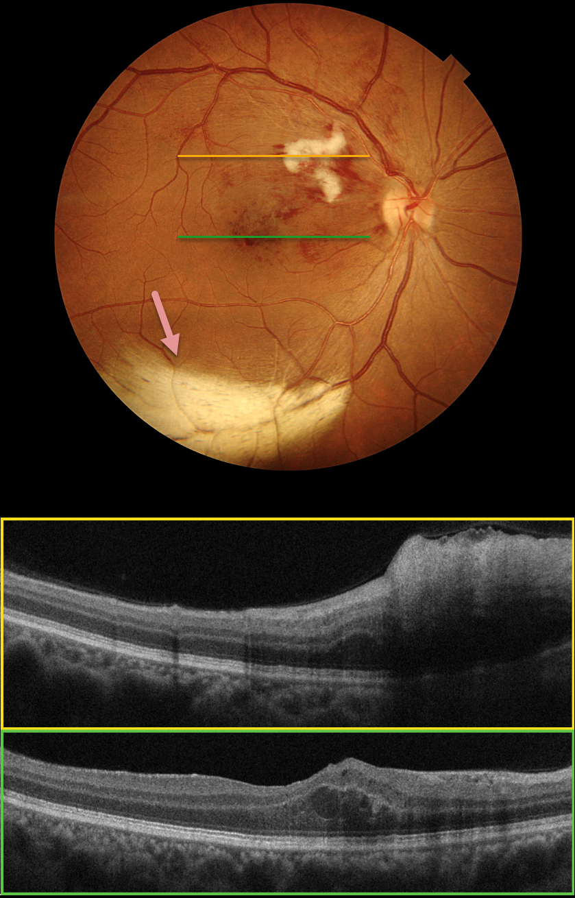
Color photography of this 48YO female’s right eye shows a major superotemporal branch retinal vein occlusion (BRVO) with foveal blood. A prominent nerve fiber layer infarct (cotton-wool spot, CWS) is present in the superonasal macula. OCT scanning shows diffuse hyperreflective inner retinal thickening through the CWS and some central foveal fluid in the outer nuclear layer. An unrelated patch of myelinated nerve fiber layer (NFL) is noted along the inferotemporal arcade. The macular edema resolved following 3 monthly intravitreal Lucentis injections (not shown).
Our patient’s eye shows two different pathologies that each involve the NFL. Myelinated NFL occurs when retinal nerve fibers develop a myelin sheath, which usually stops posterior to the lamina cribrosa. The white myelin beautifully displays the normally transparent NFL. Myelination may represent an oligodendrocytic choristoma (see Rao et al Retina 2019;39:1125-1132). Nerve fiber layer infarcts cause a more fluffy, white ischemic opacification of the NFL and are a relatively nonspecific finding in numerous disorders, including diabetes, retinal venous occlusion, hypertension, and HIV/AIDS.



