

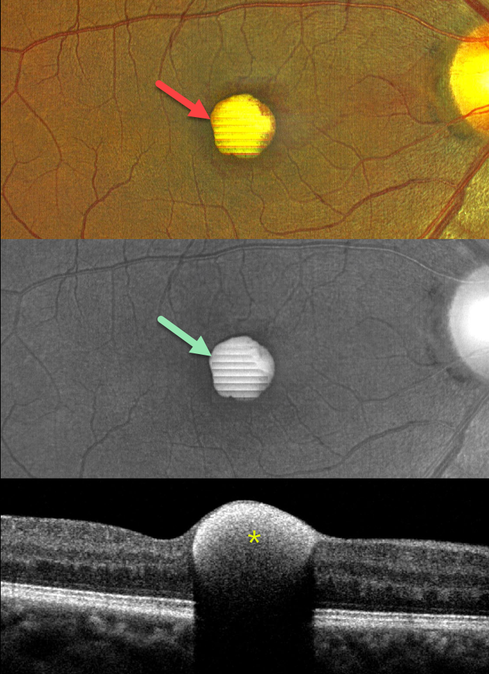
This 43YO male presented with a 1-week history of sudden decreased vision in his right eye. He denied any Valsalva maneuver. Vision was 20/70 in his right eye and 20/30 in his left eye.
Color imaging shows a foveal yellow sub-internal limiting membrane (ILM) hemorrhage. Fundus autofluorescence (FAF) shows intense hyper-FAF. Spectral domain OCT shows a hyperreflective sub-ILM lesion that shadows posteriorly.
Our patient was treated with observation, and the blood levels were greatly decreased 3 weeks later (not shown).
Learning Points:
Valsalva retinopathy was first described by Thomas Duane (Trans Am Ophthalmol Soc 1972; 70:298-313). It is characterized by usually unilateral retinal and preretinal blood caused by raised intrathoracic or intra-abdominal pressure. Various causes include coughing, heavy lifting, and vomiting. However, as in our case, sometimes patients present with these findings, denying any Valsalva maneuver (Forshaw et al, Acta Ophthalmologica 2024;102:122-123)
These hemorrhages virtually always resolve without sequelae, although the preretinal blood in severe cases can be drained into the inferior vitreous by creating a small ILM opening using either a thermal or Nd:YAG laser (Durukan et al, Eye 2008;22:214-218).
Porphyrins are organic molecules that are part of the heme molecule in hemoglobin. When these compounds break down, the devitalized blood appears yellow and becomes intensely hyperautofluorescent (Bloom and Spaide, Retinal Cases & Brief Reports 2022;16:401-402).



