

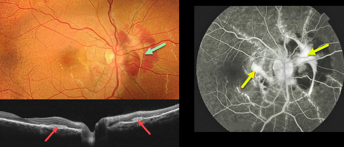
This healthy 18YO female presented with recent vision loss in her right eye. Vision was 20/40 OD and 20/30 OS.
Fundus photography shows angioid streaks extending around and radiating outwards from each optic nerve. The posterior pole has a diffuse orange skin (peaux d’orange) appearance. A fibrosed macular neovascularization (MNV) encircles the right nerve and extends into the nasal macula.
On OCT scanning, the hyperreflective MNV is located above the RPE (type 2 lesion) and is associated with minimal exudation. The MNV stains on fluorescein angiography. Intravitreal Lucentis therapy was started in her right eye.
Learning Points:
Angioid streaks are orange-yellow, irregular, crack-like dehiscences in Bruch’s membrane associated with atrophic degeneration of the overlying RPE. The orange skin (peaux d’orange) appearance is caused by a calcified Bruch’s membrane giving a yellow mottled ‘coquille d’oeuf’ (eggshell) appearance that contrasts with the normal orange RPE and choroidal pigmentation (Spaide, Retina 2015;35:392-397). These patients are at significant risk for MNV, as occurred in our patient’s right eye.
Angioid streaks are often missed clinically because their color and orientation blend with the overlying retinal vessels. Individuals with the extensive streaks and peaux d’orange, as seen in our patient, are pathognomonic for PXE, although less extensive streaks can also be seen in sickle cell and age-related macular degeneration (Eye 2023;37:1596-1601).
Our patient gave no family history of PXE. Genetic testing was not possible due to financial reasons, but we will refer her to dermatology for a skin biopsy to definitively establish this diagnosis.



