

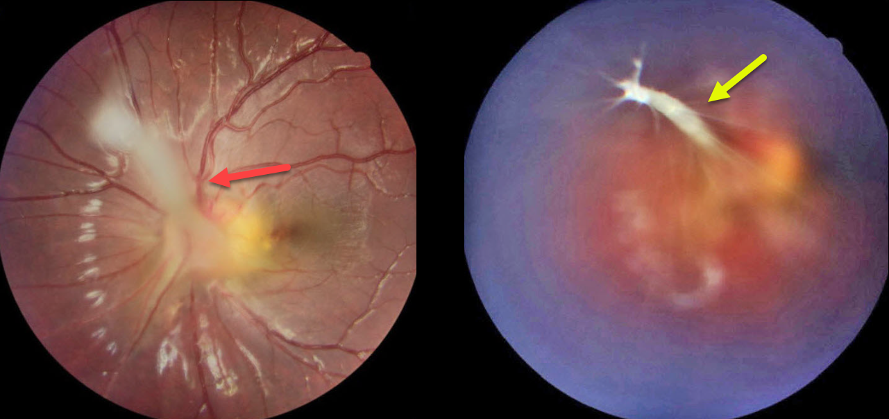
This 7YO girl presented with blurred vision in her esotropic left eye, which had previously been treated with patching of her normal right eye. Vision was 20/20 OD and 20/50 OS. Refraction was +0.25 +0.25 x180 OD and +1.00-1.50 x60 OS.
Color imaging of her left eye shows a fibrotic stalk that extends from the optic nerve head superonasally, terminating in the mid-vitreous in a spider web configuration. The macula is dragged towards the nerve, causing a shallow nasal foveal traction detachment. Lens-sparing vitrectomy will be performed in the near future.
Learning Points:
Persistent fetal vasculature (PFV), previously recognized as persistent hyperplastic primary vitreous (PHPV), represents a failure of regression of fetal vessels. It is a common cause of leukocoria and subsequent amblyopia. There is no identifiable genetic association.
Characteristically, the affected eye is microphthalmic with a shallow chamber predisposed to secondary angle closure glaucoma, a varying degree of cataract, and retrolental and/or posterior fibrovascular membranes that can cause retinal traction. If diagnosed early in life, PFV can be surgically treated with a combined lensectomy and vitrectomy. Anterior-type PFV is associated with better vision than combined or posterior types, and younger age at presentation correlates with improved vision after treatment.



