

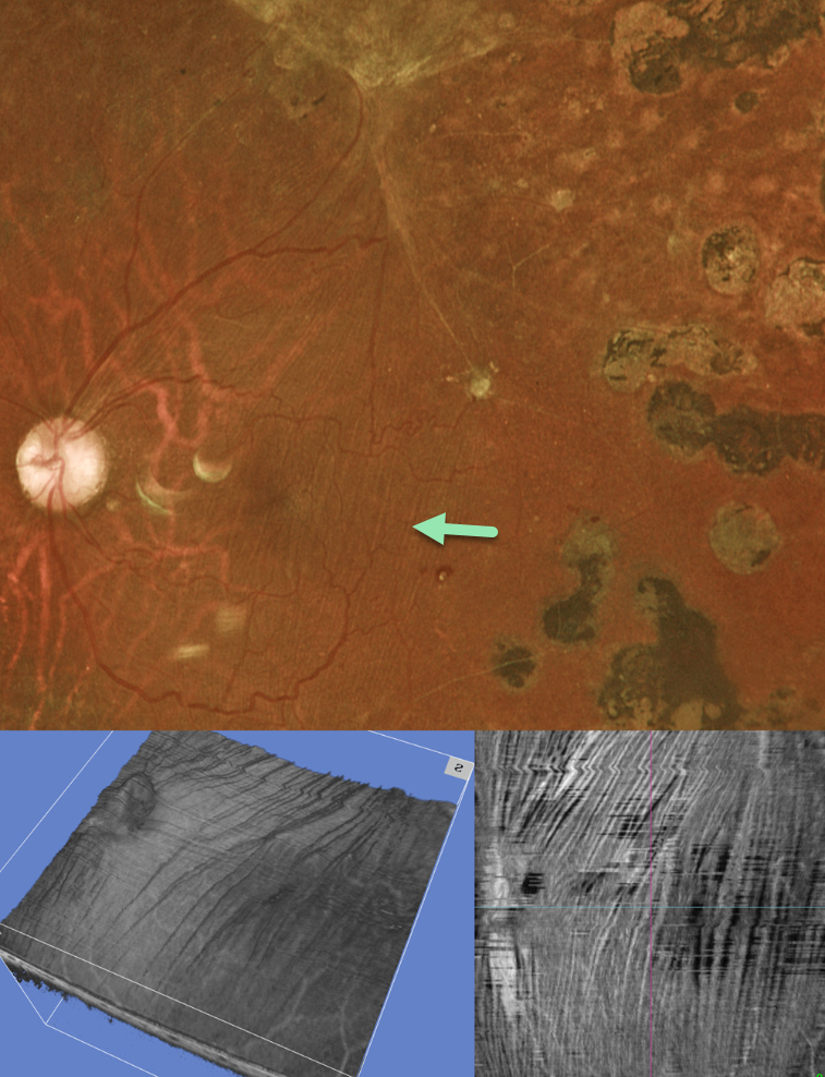
This 35YO diabetic male underwent prior vitrectomy surgery for a diabetic traction retinal detachment (TRD) five years earlier. Vision was stable at 20/40.
Optos color RG imaging shows fine macular striae extending superiorly towards the area of prior traction. Triton swept-source OCT 3D reconstruction and inner retinal en face show differing perspectives of these inner retinal folds.
Inner retinal folds are common following vitrectomy for diabetic TRD. They often fail to completely resolve, and vision can be quite good despite the persisting findings.



