

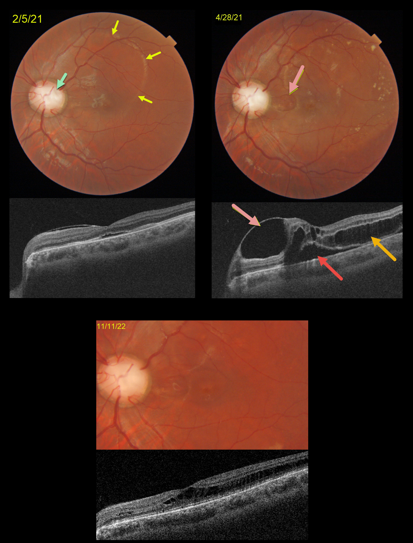
This is a follow-up to our 2/5/21 and 4/28/21 posts of a patient with bilateral optic nerve coloboma with optic pit maculopathy in his left eye.
On 2/5/21, vision was 20/70 with an optic pit and macular fluid. Despite the mostly peripheral fluid, the macular center was normal except for a trace amount of subretinal fluid (SRF).
Two months later, the peripheral fluid was better, but the central OCT findings were markedly different, with a nasal inner cyst, temporal outer schisis, and increased SRF. Although vision had decreased to 20/200, he subjectively felt his vision was better and elected continued observation.
When examined 1.5 years later on 11/11/22, the OCT findings had spontaneously improved. Temporal outer retinal schisis extends through the macular center, which is now free of SRF. Vision was 20/100, and he remained visually asymptomatic.
Learning Points:
An optic nerve coloboma can be a unilateral or bilateral condition caused by incomplete closure of the embryonic fissure.
While most optic pits are asymptomatic, they can cause vision loss due to fluid being forced into the inner retina, outer retina, and finally the subretinal space (optic pit maculopathy). The source of the fluid continues to be a point of ongoing debate, coming from either the vitreous or subarachnoid space.
A host of treatments for optic pit maculopathy have been suggested over the years. However, a recent meta-analysis suggested that a plain old vanilla vitrectomy without gas tamponade is as successful as any other procedure (Zheng et al Ophthalmology Retina 2020;4:289-299).
Our case illustrates how this fluid can spontaneously wax and wane, and that sometimes simple observation is the best option for some patients.



