

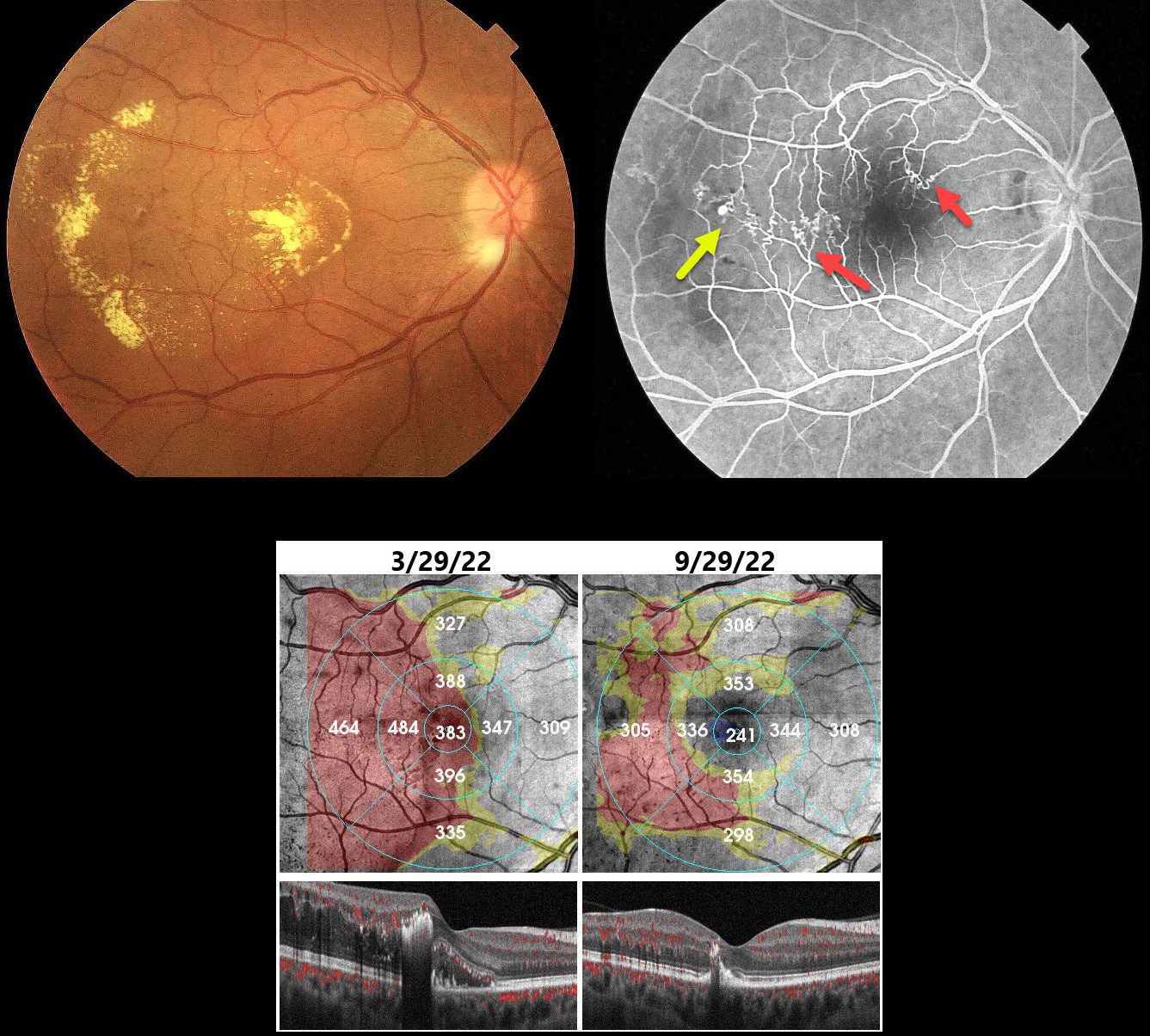
This 40YO hypertensive male presented with gradual vision loss in his right eye. Vision was 20/200 OD and 20/20 in his normal OS.
Color imaging shows central and temporal macular lipid and telangiectasia. Fundus fluorescein angiography shows a superotemporal branch retinal vein occlusion (BRVO) with superonasal and temporal collateral vessels. A retinal venous macroaneurysm (RVM) is noted distally.
Our patient was initially treated unsuccessfully with several monthly Lucentis injections. The macular edema and lipid improved following macular grid laser with focal treatment of the RVM.
Learning Points:
In 1990 Cousins et al reported that RVMs can be an additional finding in occlusive venous disease, always in association with collateral vessels or capillary or arterial macroaneurysms (AJO 1990;109:567-570). These vascular abnormalities tend to have a higher rate of intraretinal lipid (40%) but are less likely to affect central vision since they are usually outside the macula. They tend to be associated with retinal ischemia, thus increasing the risk of developing retinal neovascularization (32%).



