

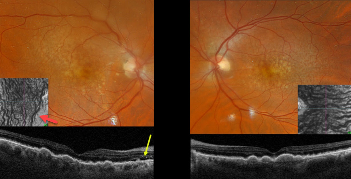
This 77YO male was referred for asymptomatic neovascular AMD in his right eye. Vision was 20/50 bilaterally.
Optos color RGB imaging shows extensive bilateral larger drusen and subretinal drusenoid deposits, which are evident on Triton swept-source OCT. Localized subretinal fluid is noted just temporal to the right nerve.
Although the choroid is not particularly thickened, it is relatively thick for a 77YO, and large vessels span the full-thickness choroid. En face choroidal OCT shows markedly abnormal dilated vessels.
Learning Points:
Originally described by Phasukkijwatana et al in 2018 (Retina 2018;38:1652-1667), peripapillary pachychoroid syndrome presents with nasal macular intraretinal and subretinal fluid. These eyes can look very similar to those of patients with optic pit maculopathy, but without the pit. Additional findings include a thicker nasal than temporal macular choroid, choroidal folds (75% of cases), shorter axial lengths (<23mm), and hyperopia. There is no angiographic leakage despite the presence of OCT fluid, which often spontaneously waxes and wanes. Symptomatic fluid can successfully be treated with photodynamic therapy.
We assume our patient’s fluid is related to his pachychoroid disease rather than nAMD. Observation was recommended.



