

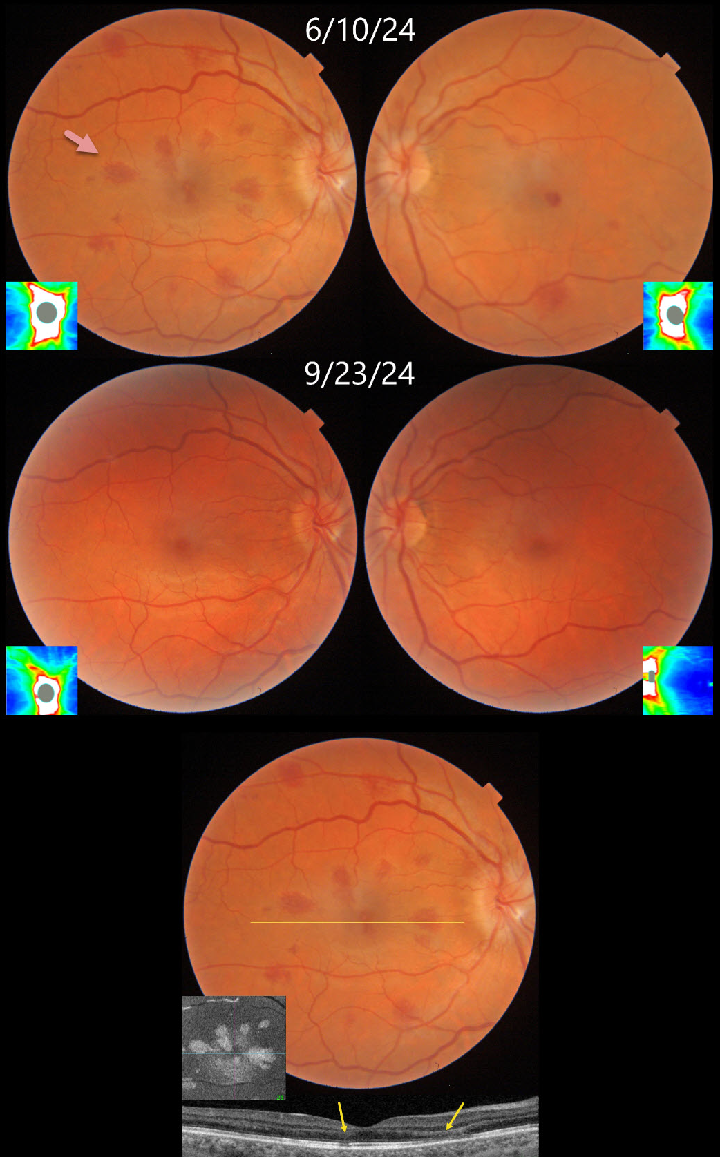
This 34YO female underwent uterine ablation 2 weeks earlier for heavy menstruation and severe secondary iron deficiency anemia. Although we don’t know what her hemoglobin level was prior to the procedure, it had risen to 10.7 postoperatively. Vision was 20/30 OD and 20/50 OS.
Triton color photography shows bilateral teardrop-shaped macular outer plexiform layer (OPL) hemorrhages, with greater involvement in her right eye. Numerous more peripheral deep and white-centered hemorrhages are also noted. Swept-source OCT confirms the location of the macular blood. En face OCT shows a more dramatic view of their petaloid shape. There is also mild optic nerve edema, especially in the right eye. The hemorrhages and nerve swelling mostly resolved with iron supplementation several months later.
Learning Points:
Anemia-related retinal findings most commonly include hemorrhages in the retina and sub-internal limiting membrane. Severe anemia is a rare cause for optic nerve swelling and should always be considered in patients with atypical idiopathic intracranial hypertension (Biousse et al, AJO 2003;135:437-446). The underlying pathogenesis of anemia-induced optic nerve swelling remains unknown.
Radiating deep macular hemorrhages are seen in multiple conditions, including Valsalva and trauma (Baumal et al, Br J Ophthalmol 2021;105:374-380). We have not previously seen these hemorrhages in isolated anemia. Their unique radiating appearance is caused by blood accumulating in Henle’s layer. Kon Graversen et al described hemorrhagic unilateral retinopathy, an idiopathic disorder with a similar appearance (Retina 2014;34:483-489).



