

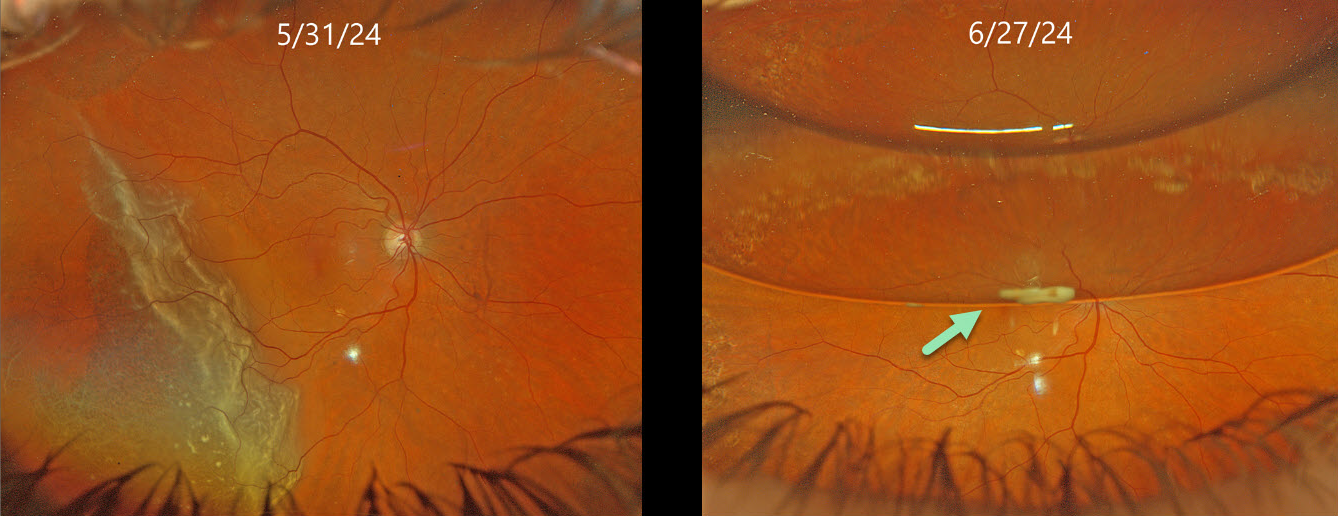
This 72YO female presented with one day of peripheral vision loss in her right eye. Vision was 20/25 OD and 20/25 in her healthy OS.
Optos color RG imaging (image 2, top) shows a temporal macula-on rhegmatogenous retinal detachment (RRD). Pars plana vitrectomy (PPV) with a C3F8 bubble was performed later that day. On her 1-month postoperative visit, vision was 20/50 with about a 50% gas bubble. A greyish globular deposit was adherent to the inferior edge of the gas meniscus.
Learning Points:
Russell et al recently reported a unique ophthalmoscopic sign, a meniscus micropyon, that follows PPV with gas tamponade for all causes (Retina 2024;44:1766-1776). Features they noted that distinguish this sterile micropyon from post-PPV fibrin include delayed appearance, hyperautofluorescence, and absence of fibrinous strands or sheets in the anterior chamber or vitreous. Vitreous biopsy in two of their cases revealed predominantly white blood cells. The micropyon appearance was associated with epiretinal proliferation (80%). Recurrent RRD from proliferative vitreoretinopathy (PVR) developed in 33% of eyes, 12% re-detached without frank PVR, and 18% developed worsening epiretinal membranes.
Fortunately, our patient’s postoperative course was uneventful. The micropyon resolved at the following visit and 4 months postoperatively, vision was 20/20 and the retina remained completely attached without epiretinal proliferation (not shown). Since the publication of Russell’s article, we have seen several patients with this biomarker, so we suspect it to be a not uncommon finding.



