

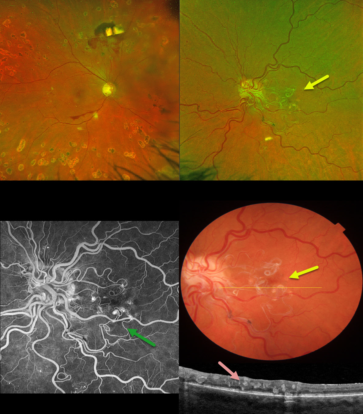
This 53YO female with a history of type 2 diabetes was referred for a symptomatic vitreous hemorrhage in her right eye from proliferative diabetic retinopathy. She received prior panretinal photocoagulation (PRP) in this eye elsewhere. Vision was 20/60 OD and 20/70 OS.
Optos color RG imaging OD shows preretinal blood in the inferior macula, and PRP scarring is noted superior to the nerve. The left eye shows a tangle of dilated and partially sheathed vessels extending from the nerve into the central macula. The more peripherally located vessels are dilated and tortuous.
On B-scan OCT, these vessels are noted in all retinal layers. These vessels did not leak on fluorescein angiography. Neurologic imaging was negative.
Learning Points:
A retinal arteriovenous malformation (AVM) is a congenital connection between an artery and a vein without an intervening capillary network. These lesions appear along a spectrum ranging from small, isolated lesions to the large ‘bag of worms’ appearance as seen in our patient. These more extensive lesions are usually part of the Wyburn-Mason Syndrome, which includes ocular and midbrain vascular lesions. Our patient has an isolated retinal AVM, as there are no intracranial lesions.
Unlike diabetic neovascularization that proliferates from the retinal surface into the cortical vitreous, the vessels in an AVM are intraretinal. Even though the AVM somewhat mimics diabetic disc neovascularization, its presence in our diabetic patient is purely coincidental. Although retinal AVMs are usually stable lesions, retinal hemorrhages, vitreous hemorrhage, and venous occlusions may occur.



