

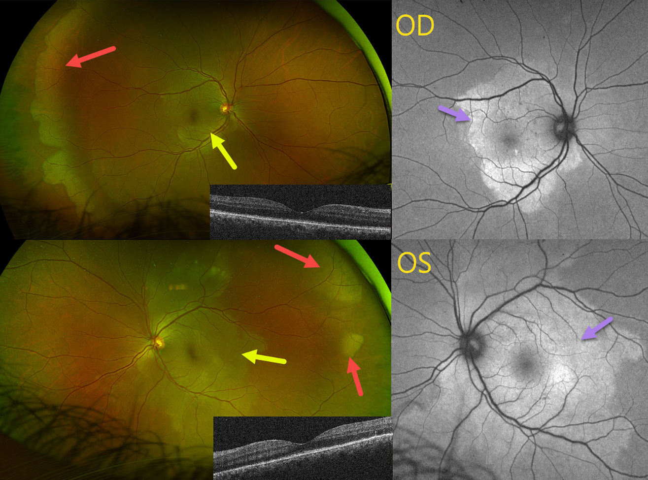
This 42YO female presented with 1 month of bilateral blurred vision. Vision was 20/30 OU.
Optos color RG imaging shows bilateral peripheral and posterior white areas without pressure. Correct??
WRONG! These posterior lesions are hyperautofluorescent. Macular OCT scanning shows variable loss of the outer retinal bands. Syphilis testing was positive, and she was immediately referred to an infectious disease specialist for treatment. When she returned 4 months later following a full course of parenteral penicillin, vision was 20/40 OD, 20/20 OS, with normalization of all fundus findings (not shown).
Learning Points:
Ocular manifestations of syphilis vary widely and include virtually all variations of anterior and posterior segment inflammation. Originally described by Gass (Ophthalmology 1990;97:1288-1297), placoid chorioretinitis is one of the more common presentations. Inflammation is localized to the outer retina and RPE and appears on OCT as disruption of the outer retinal bands and hyperreflective pyramidal lesions as seen in our patient (Hu et al, Ophthalmology Retina 2022;6:172-178). The outer retinal damage unmasks the underlying RPE, accounting for the hyperfluorescent placoid lesions.



