

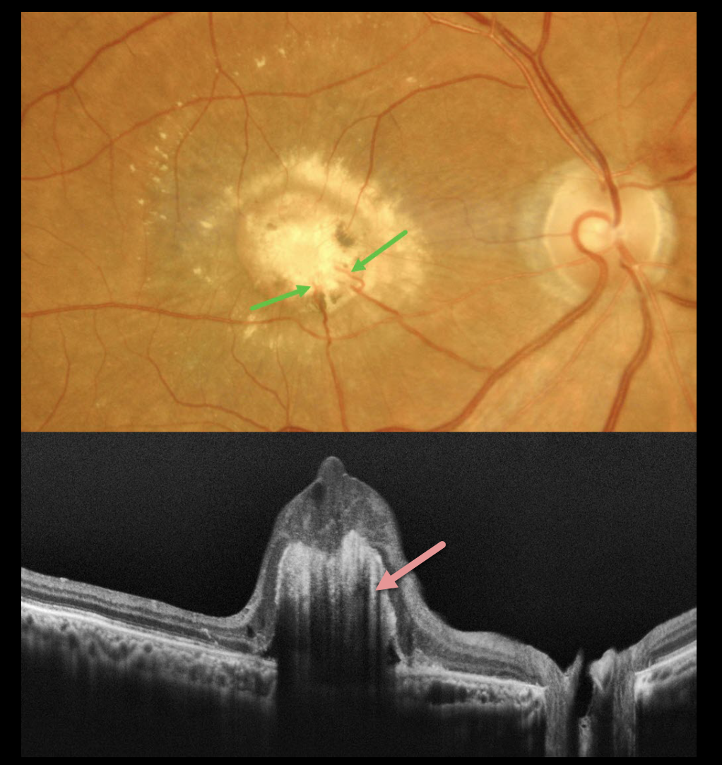
This healthy 64YO male presented with gradual vision loss in his right eye. Vision was 20/2000 OD and 20/20 in his normal OS.
Color imaging shows a yellow-white subfoveal nodular lesion with surrounding subretinal lipid. Several retinal choroidal anastomoses are noted within the lesion inferiorly. OCT scanning shows a hyperreflective elevated deep retinal lesion with overlying disorganized retina. There is marked posterior shadowing obscuring all deep retinal and choroidal detail.
Learning Points:
Choroidal granulomas usually appear as solitary, creamy white, round subretinal lesions. They are generally hyporeflective on OCT imaging (Invernizzi et al, Retina 2015;35:525-531) and can extend into the subretinal space, as in our patient. Causes include sarcoidosis, tuberculosis, Bartonella, and VKH.
Under normal conditions, the retinal and choroidal circulations remain separate and distinct. Rarely, the two circulations directly connect via a chorioretinal anastomosis. These can occur in chorioretinal scars, in macular telangiectasia, and in disciform scars.
Our best guess is that this lesion represents a vascularized choroidal granuloma. Due to its chronicity and lack of significant overlying retinal exudation, observation was recommended.



