

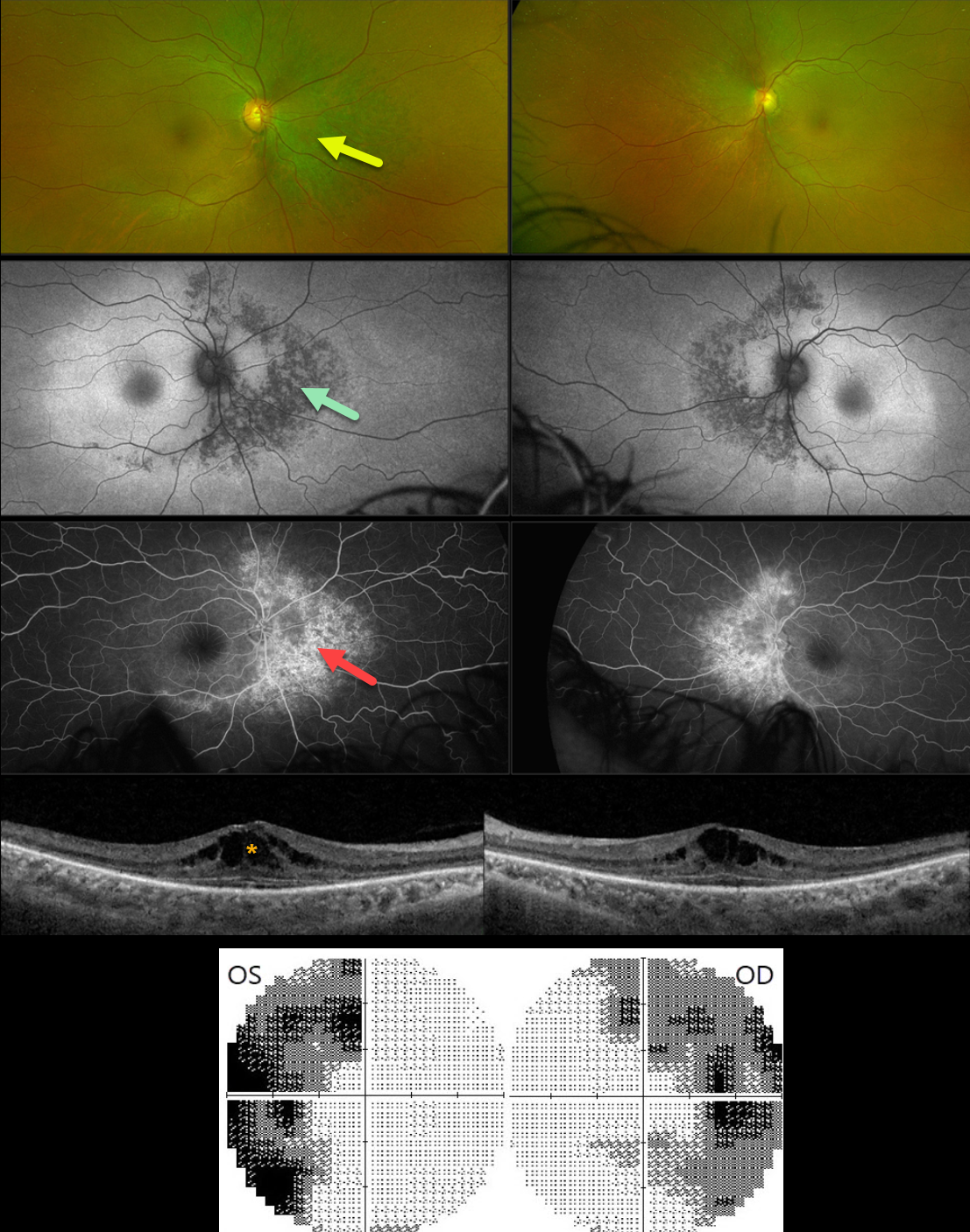
This 54YO female was referred for asymptomatic retinal changes. Vision was 20/30 OD and 20/20 OS.
Visual fields showed bitemporal hemianopia. What is your diagnosis?
Optos color RG imaging shows pigmentary changes with intraretinal pigment migration extending superiorly, nasally, and inferiorly from the optic nerves. These changes are hypoautofluorescent and appear as angiographic window defects. Bilateral cystoid edema is seen on Triton swept-source OCT.
Learning Points:
Although bitemporal hemianopia usually signifies retrobulbar pathology at the optic chiasm, retinal disease can sometimes also cause similar field loss. Our patient’s retinitis pigmentosa (RP) changes were centered around each optic nerve, causing the hemianopia. Genetic testing revealed double heterozygous pathogenic ARSG mutations. This genotype has been associated with atypical Usher syndrome (Fowler et al, Ophthalmic Genetics 2021;42:338-343).



