

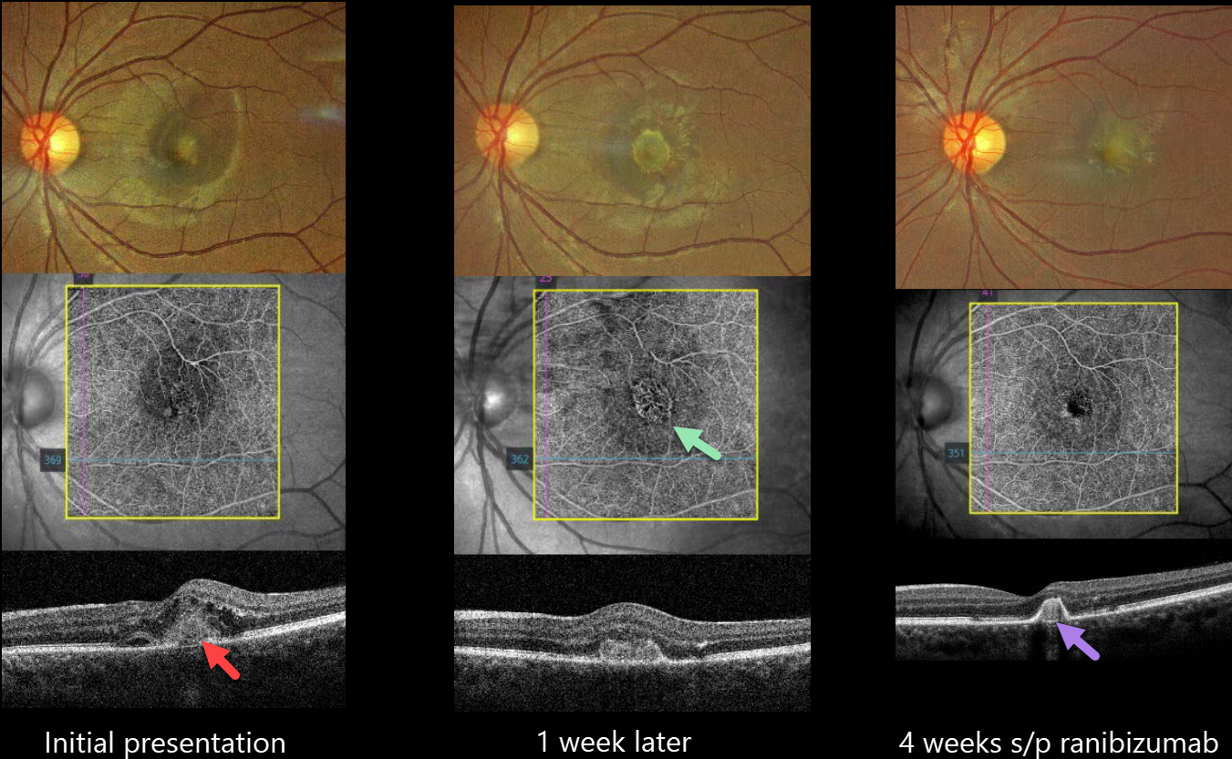
This 20YO female presented with 2 weeks of decreased vision in her left eye. Vision was 20/25 in her normal right eye and 20/60 in her left eye.
Color imaging shows foveal fluid with an amelanotic subfoveal lesion. OCT angiography (OCTA) shows a small subfoveal macular neovascularisation (MNV). An OCT B-scan shows a type 2 MNV (above the RPE) with intraretinal fluid and numerous hyperreflective foci.
Although we recommended an intravitreal ranibizumab biosimilar that day, she refused treatment and returned one week later with 20/200 vision and marked enlargement of the MNV on OCTA.
The injection was given, and 4 weeks later, vision improved to 20/40, with a dramatic treatment response characterized by decreased size and exudation.
Learning Points:
Idiopathic MNV in younger patients was first described by Cleasby in 1976 (AJO 1976;81:590-596). They tend to be type 2 lesions and have a better natural history than typical neovascular AMD (Ophthalmology 1998;105:1816-1820). There is usually an excellent response to anti-VEGF therapy (Kodjikian et al, Retina 2022;42:290-297), as in our patient who initially had both a dramatic rapid enlargement of the untreated lesion, followed by an equally rapid treatment response.



