

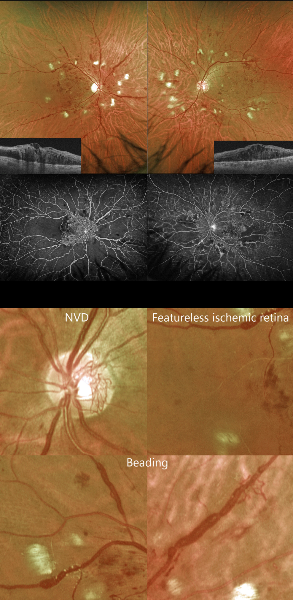
This 41YO male with type 2 diabetes complained of vision loss for 5 months. He was recently hospitalized for severe systemic hypertension. Vision was counting fingers OD and 20/60 OS.
Optos color RG imaging shows bilateral venous beading and extensive cotton wool spots. Diffuse central edema is noted on Triton swept-source OCT, and severe ischemia is seen on fluorescein angiography.
But on closer inspection, the severe ischemia is evident clinically. The retina between the larger vessels is featureless, and many of the veins are sausaged and beaded. Once these clinical features are recognized, the angiographic findings can usually be predicted.
There is a relative lack of severe retinal hemorrhages and neovascularization (only disc neovascularization is present). The extensive cotton wool spots are more hypertensive than diabetic changes but indicate ischemia of the nerve fiber layer.
The patient subsequently underwent full bilateral panretinal photocoagulation and anti-VEGF therapy.



