

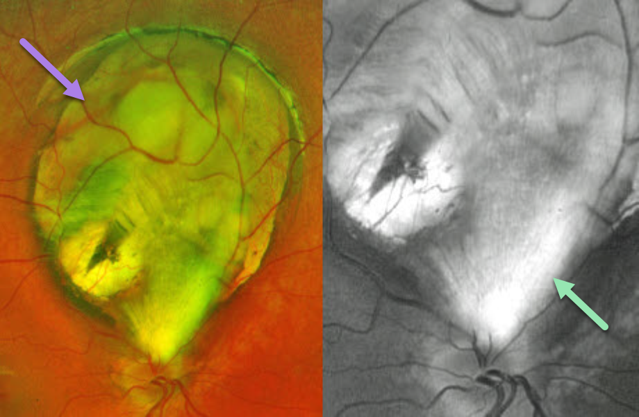
This healthy 8YO girl was referred for an asymptomatic retinal finding in her right eye. Vision was 20/30 in her right eye and 20/30 in her left eye. Anterior segments were normal.
Optos color RG imaging shows a superior choroidal coloboma that almost extends to the disc margin. An incidental patch of myelinated nerve fiber layer (NFL), best imaged in the green channel, extends from the superior nerve into the inferior aspect of the coloboma.
Learning Points:
Choroidal colobomas are caused by failure of the optic vesicle and choroidal fissure to close during fetal development. Choroidal coloboma can present as solitary or multiple lesions. Visual prognosis depends on the location and involvement of the optic nerve and macula. We will follow our patient yearly due to her increased risk of retinal detachment.
Myelinated NFL is a relatively uncommon and usually isolated finding present at birth. Most patients are asymptomatic, although some may have a corresponding relative scotoma. Myelination can progress in about 10% of cases and can also resolve following retinal ischemic events. Myelinated NFL occurs when retinal nerve fibers develop a myelin sheath, which usually stops posterior to the lamina cribrosa. The myelination may represent an oligodendrocytic choristoma (see Rao et al Retina 2019;39:1125-1132).



