

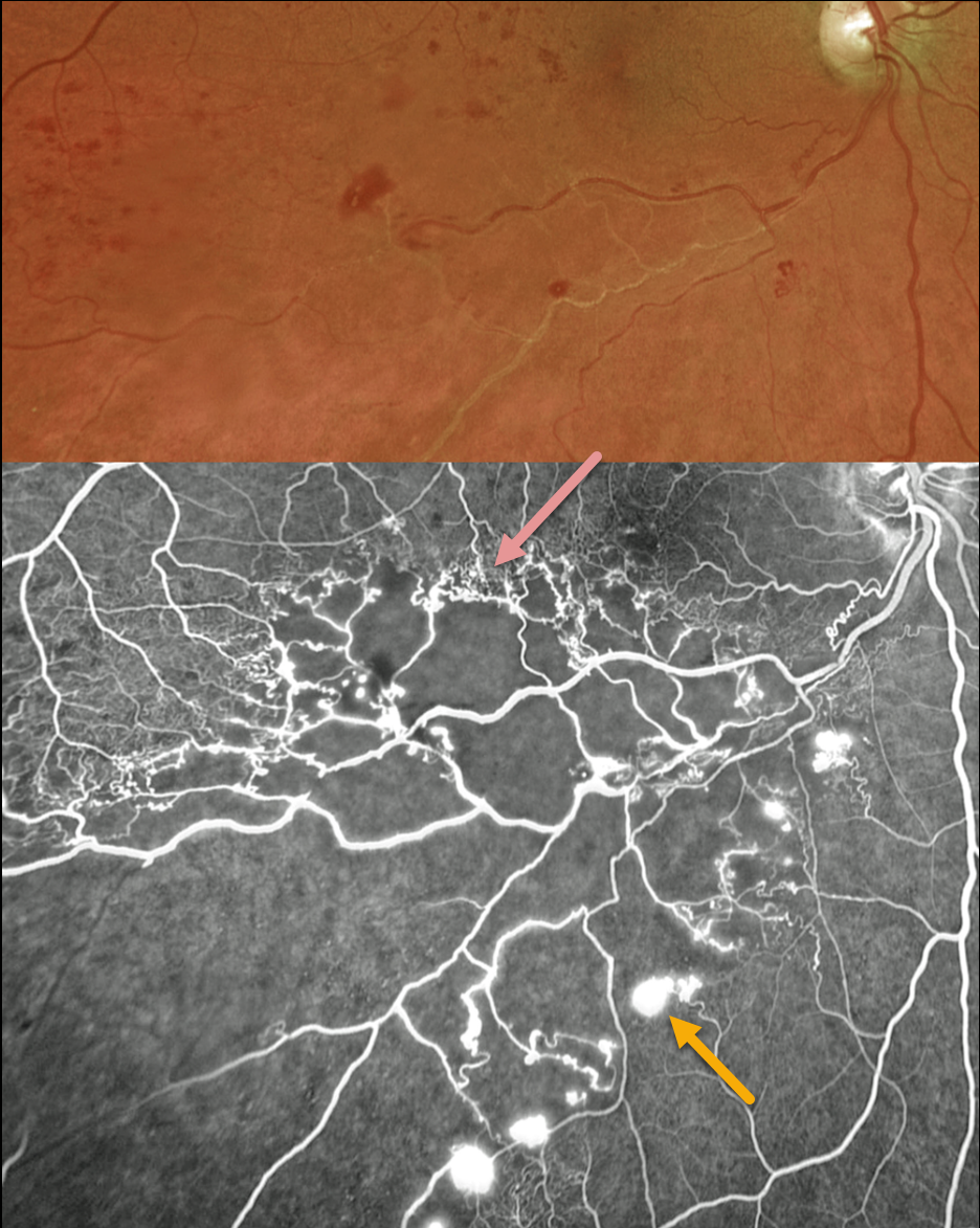
This 56YO male presented with a major inferotemporal ischemic branch retinal vein occlusion (BRVO) in his right eye and 20/40 vision. Despite the deceptively good vision, Optos ultra-widefield imaging showed marked ischemia throughout the distribution of the occlusion, which fortunately spared the central macula. A row of collateral vessels extends along the superior edge of the occlusion, with retinal neovascularization extending along its inferior extent. Leaking disc neovascularization is also present. A scatter laser throughout the area of ischemia was performed, but unfortunately, he was then lost to follow-up.
Learning Points:
In 1986, the Branch Vein Occlusion Study Group reported that scatter laser decreased the risk of vitreous hemorrhage from retinal neovascularization in BRVO, and suggested that laser should be applied once neovascularization develops (Arch Ophthalmol 1986;104:34-41). Thirty-five years later, scatter laser remains the gold standard, although some may prefer to first stabilize neovascularization with anti-VEGF therapy before laser.
Collateral vessels are dilated, pre-existing vessels that bypass an occlusion. These are most commonly seen at the posterior pole following retinal venous occlusions and are exceedingly rare with arterial occlusions. The endothelial tight junctions are intact, so they don’t leak angiographically. This helps differentiate them from neovascularization, which does leak.



