

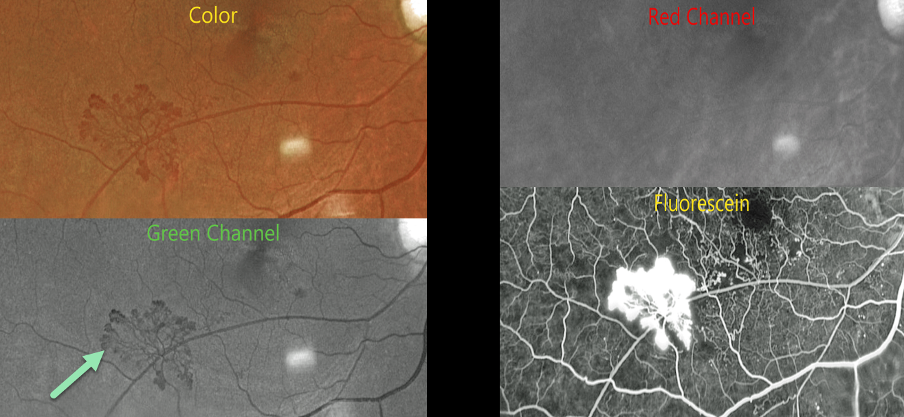
This 54YO male with proliferative diabetic retinopathy (PDR) and retinal neovascularization was evaluated with Optos multimodal imaging.
Optos color RG imaging shows active neovascularization in the inferotemporal macula.
The neovascularization is best seen in the green channel, which isolates the RPE and neurosensory retina. The blood vessels virtually disappear with the red channel, which is best for viewing the choroid. Finally, the vessels profusely leak on fluorescein angiography.
Learning Points:
Optos RG color images are generated from a red (635nm) and green (532nm) laser. The absence of a blue channel results in their greenish tint, which, at first, many clinicians find somewhat unsettling. However, much information is contained in these images once one understands how they are constructed. As shown in our patient, these images allow isolation of the neurosensory retina/RPE and choroid and often reveal more information than is visible in a true-color image.
Red neovascularization on an orange background (color image) is much more difficult to see than black neovascularization on a grey background (green channel). Recently, Optos introduced a blue channel to its California model, which, in our opinion, now offers the best of all worlds: stunning true-color images along with the ability to view each channel separately.



