

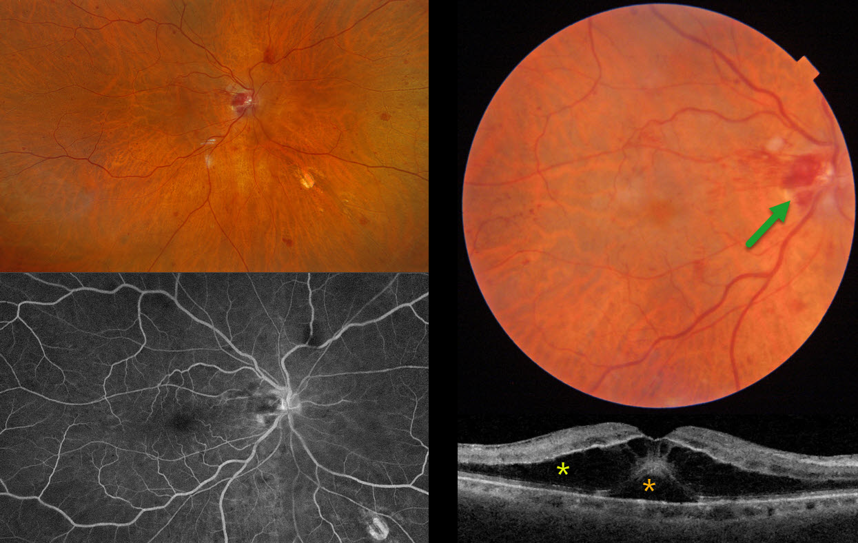
This 81YO male presented with two days of vision loss OD. He has a history of diet-controlled type 2 diabetes and a 5-year history of multiple myeloma. He was examined by us 2 weeks earlier for a routine visit for inactive herpes simplex iridocyclitis. Vision was 20/40 on that visit with a normal OCT (not shown), but had decreased to counting fingers at the current visit.
Triton color imaging shows a faint, round serous macular detachment with scattered small inner retinal and nerve hemorrhages. Swept-source OCT shows subfoveal fluid with overlying cystic edema in the outer nuclear layer. Optos color RGB imaging shows scattered retinal hemorrhages throughout the right posterior pole, with a normal left fundus. Fluorescein angiography shows no leakage.
Learning Points:
Monoclonal gammopathies (paraproteinemia) result from the production of monoclonal gamma-globulins in the blood, including multiple myeloma (IgG or IgA) and Waldenstrom macroglobulinemia (IgM). Retinal findings are often caused by hyperviscosity (Rajagopal et al, Surv Ophthalmology 2016;61:236-247) and include intraretinal or deep retinal hemorrhages, dilated vessels, or vascular occlusions.
A unique retinopathy found in these disorders is termed paraproteinemic maculopathy (Mansour et al, Ophthalmology 2014;121:1925-1932). Somewhat simulating optic pit maculopathy, patients develop serous macular detachments with overlying cystic outer retinal edema and occasionally outer macular holes. There is no angiographic leakage. The macular fluid, which is usually refractory to laser photocoagulation, intravitreal injections, and photodynamic therapy, is treated primarily by decreasing blood immunoglobulin levels.
Unfortunately, our patient died 2 months after our examination.



