

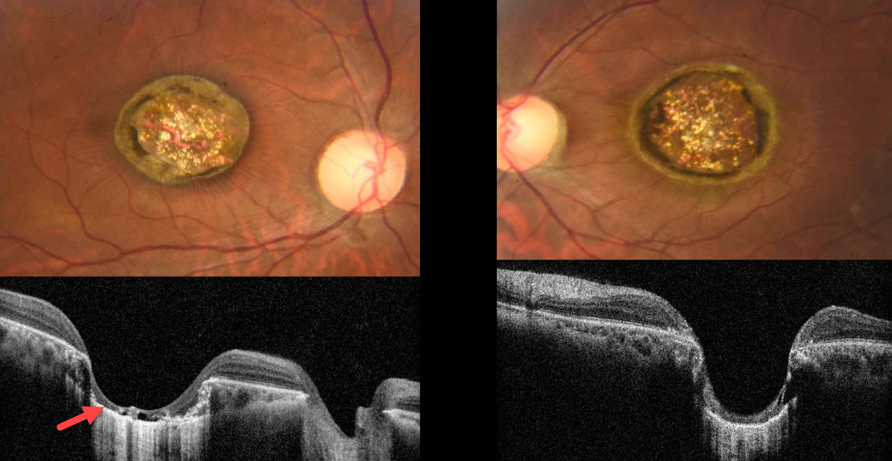
This healthy 21YO male presented with a lifelong history of stable bilateral vision loss. There was no significant ocular family history, and ocular examination of family members revealed no abnormalities. Vision was 20/400 OU. There was bilateral horizontal nystagmus.
Color photography shows fairly symmetrical macular colobomas. Fine inner retinal striae radiate outwards from the coloboma OD. OCT scanning shows near-total choroidal loss within each coloboma, with a thinned, atrophic retina draped against bare sclera.
Learning Points:
Choroidal colobomas are most commonly due to failure of the optic vesicle and choroidal fissure to close during embryonic development and are usually found inferonasally. They can present as solitary or multiple lesions. Macular colobomas are most commonly either post-inflammatory (toxoplasmosis, cytomegalovirus, and Zika) or from an inherited retinal disorder (IRD), including North Carolina macular dystrophy (NCMD).
The symmetry of our patient’s lesions argues for an IRD. However, we doubt this is NCMD, given the lifelong history of poor vision and nystagmus. Due to financial constraints, the patient refused all blood work and genetic testing. It is



