

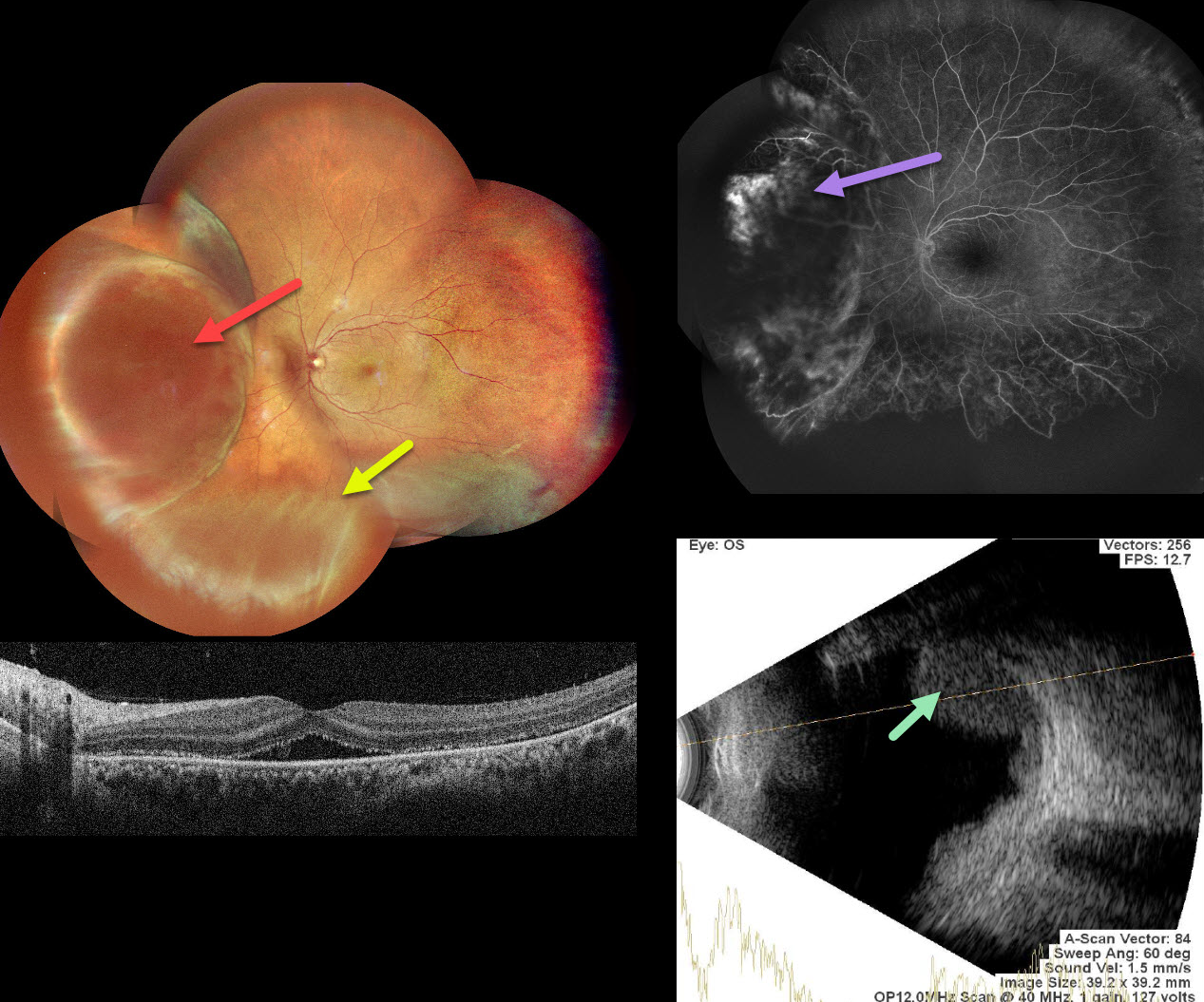
This 39 YO female presented with 1 month of decreased vision in her left eye. Vision was 20/20 in his normal OD and 20/60 OS.
Pseudocolor SLO imaging shows a large, nasal, well-circumscribed amelanotic choroidal lesion with an inferior exudative retinal detachment. OCT shows foveal subretinal fluid with likely shed outer segments along the posterior retinal surface.
Fluorescein angiography shows some leakage within the choroidal tumor. Severe peripheral retinal ischemia is noted temporally and inferiorly. The lesion shows moderate homogenous internal reflectivity on B-scan ultrasonography. MRI (not shown) was suggestive of a uveal malignant melanoma (MM), measuring 11×7 mm in size.
She was referred to ocular oncology for further treatment and evaluation.



