

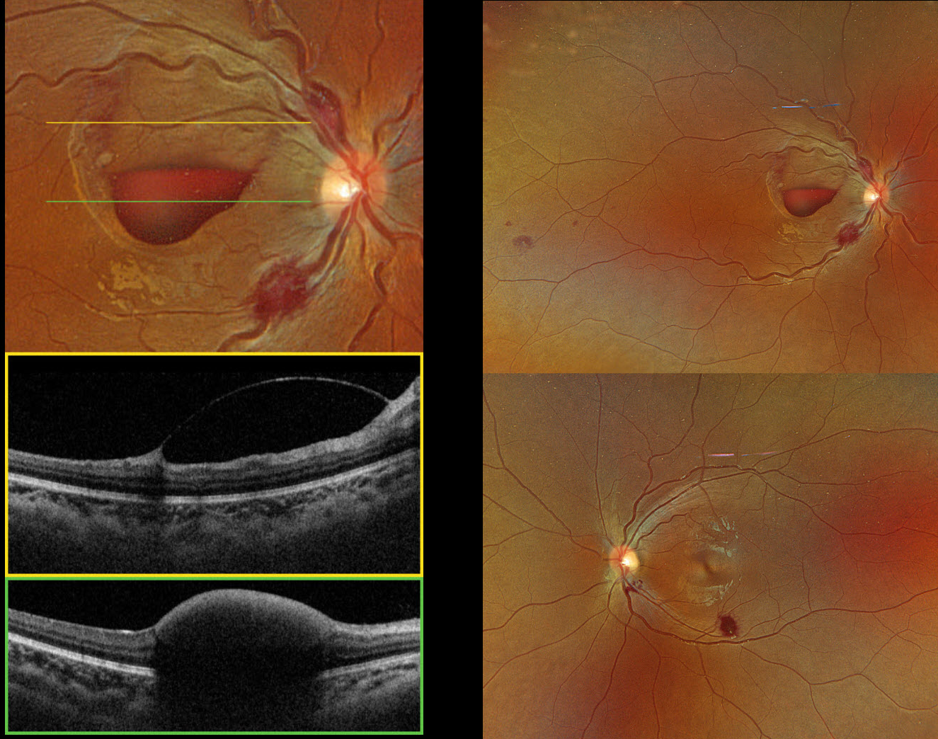
This previously healthy 14YO female presented with two weeks of vision loss in her right eye. Vision was hand motion OD and 20/25 OS.
Optos color RGB imaging shows bilateral inner and deep retinal hemorrhages, along with an inferior macular sub-internal limiting membrane (ILM) hemorrhage OD. Triton swept-source OCT confirms the location of the macular blood OD.
She was immediately sent to the emergency room. Complete blood count revealed pancytopenia with HgB 3.8, WBC 1.4, and platelets 54, and the patient was subsequently diagnosed with acute myelogenous leukemia. She was immediately lost to follow-up.
Learning Points:
Ocular leukemic manifestations are primarily caused by accompanying hematologic abnormalities, including hyperviscosity, anemia, and thrombocytopenia (Soman et al, Ophthalmology Retina 2018;2:17-23). Posterior segment findings include retinal hemorrhages (sub-ILM, nerve fiber layer, or deeper, white-centered), cotton-wool spots, dilated retinal veins, and serous retinal detachment. Leukemic infiltration of the optic nerve, retina, and choroid can also occur. Sub-ILM hemorrhages are usually self-limiting and resolve within several months of treatment of the underlying disease (Ophthalmology Retina 2018;2:494-501).



