

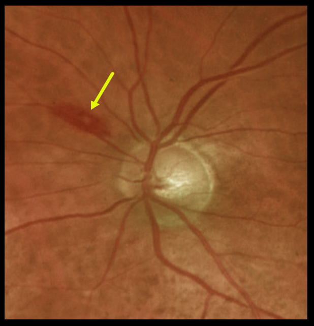
This 84YO female was found to have a white-centered retinal hemorrhage superonasal to the optic nerve disc.
Complete blood count revealed a white blood count of 19,000 and a hemoglobin of 7.9. She was subsequently diagnosed with atypical diffuse large B-cell lymphoma.
Learning Points:
The term “Roth spot” was named after Dr. Moritz Roth, who, in 1872, noted white-centered hemorrhages in patients with bacterial endocarditis.
He felt that these white spots were septic emboli that originated from an infected cardiac valve. However, similar lesions can be seen in many other disorders, including leukemia, hyperviscosity, anemia, diabetes, and hypertension.
The white material may represent a variety of causes, including fibrin. These lesions are best called white-centered hemorrhages, unless one is specifically referring to a hemorrhage in a patient with bacterial endocarditis.
For a review of white-centered retinal hemorrhages, see Duane et al Ophthalmology 1980;87:66-69.



