

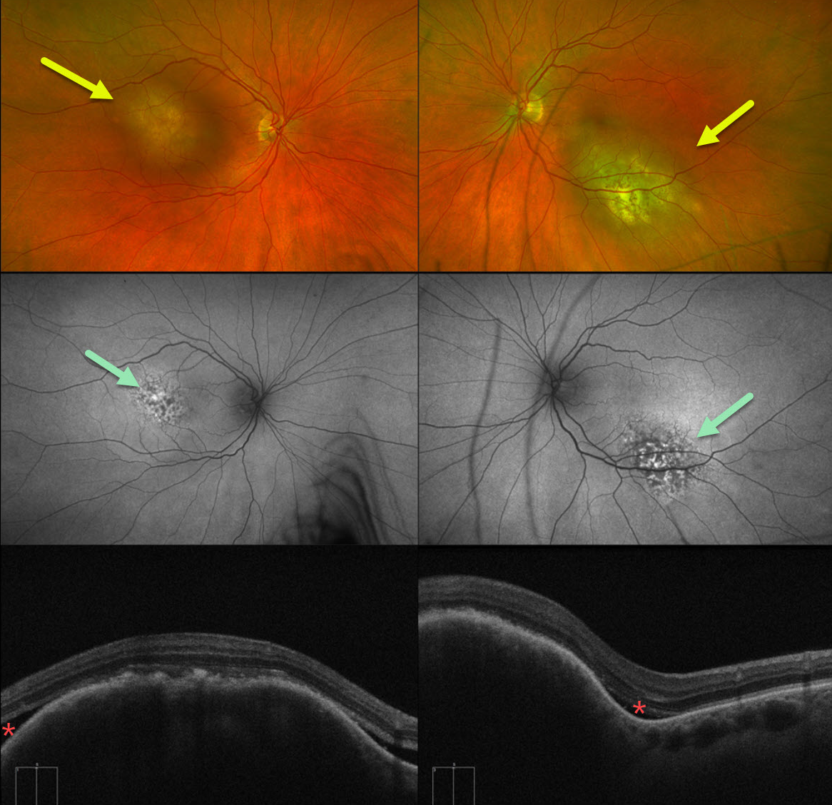
This 62YO female presented with 1 week of bilateral vision loss. She had a known history of metastatic urothelial bladder carcinoma. Vision was counting fingers OD and 20/100 OS.
Optos color RG imaging shows bilateral elevated submacular choroidal lesions. These lesions were clinically creamy in coloration, although they appeared variably amelanotic and pigmented on Optos pseudocolor imaging. Fundus autofluorescence (FAF) shows speckled hyper- and hypo-FAF overlying the superonasal portion of the lesion OD and the inferonasal portion of the lesion OS. The remainder of each lesion is mildly hyper-FAF. Some localized overlying subretinal fluid is present along the edges of the choroidal tumors.
Although uveal melanoma is the most common primary intraocular malignancy, metastatic choroidal tumors are the most common intraocular malignancies. Lung and breast cancer are the most common sources.
We immediately spoke with the patient’s oncologist and suggested referral to a radiation oncologist for external beam radiation to the choroidal metastases. Unfortunately, she died within several months of seeing us.



