

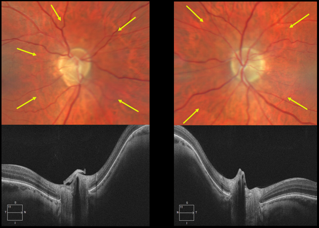
This 54YO male patient presented with asymptomatic bilateral optic nerve conus. Optos color imaging of each eye shows each optic nerve and peripapillary tissue to be sunken from the plane of the surrounding fundus. These findings are captured on OCT.
Learning Points:
An optic nerve conus is a staphylomatous-like depression of the optic nerve and surrounding tissue. This finding is usually seen in high myopia, although our patient had mild bilateral hyperopia.



