

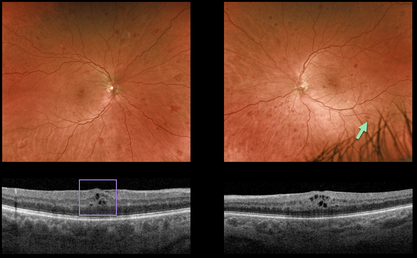
This 50YO male presented with bilateral asymptomatic retinal hemorrhages. He gave a 5-year history of well-controlled type 2 diabetes with a recent A1C of 6.3%, and was previously diagnosed with only mild nonproliferative diabetic retinopathy.
Optos imaging shows scattered retinal hemorrhages in all quadrants, most of which are deep with white centers. Mild bilateral macular edema is seen on OCT scanning.
The deeper, round retinal hemorrhages were felt to be atypical for diabetes. We therefore ordered a complete blood count (CBC), which revealed a white blood cell count over 80,000 u/L and polychromatic red blood cells.
Immediate referral to oncology revealed undiagnosed chronic myelogenous leukemia, and treatment was initiated.
Learning Points:
The term “Roth spot” was named after Dr. Moritz Roth, who, in 1872, noted white-centered hemorrhages in patients with bacterial endocarditis. He felt that these white spots were septic emboli that originated from an infected cardiac valve.
However, similar lesions can be seen in many other disorders, including leukemia, hyperviscosity, anemia, diabetes, and hypertension.
The white material may represent a variety of causes, including fibrin. So these lesions are best-called white-centered hemorrhages, unless one is specifically referring to a patient with bacterial endocarditis. See Duane et al Ophthalmology 1980;;87:66-69.



