

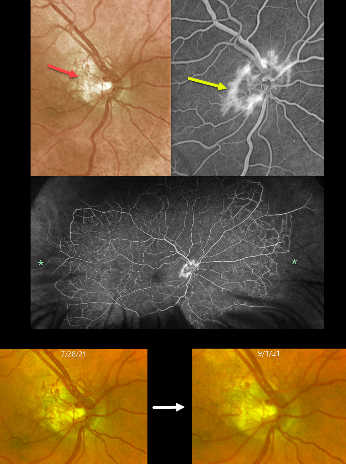
This 57YO male had a history of an old central retinal vein occlusion (CRVO) in his left eye with hand motion vision. After being lost to follow-up for 5 years, he presented on 7/28/21 with an asymptomatic CRVO with 20/25 vision in his right eye.
Optos color RG imaging shows diffuse disc neovascularization (NVD) that leaks on angiography. Ultrawidefield fluorescein angiography shows good posterior perfusion but with marked encircling peripheral ischemia. The neovascularization partially regressed 6 weeks after panretinal photocoagulation and completely regressed with subsequent laser and anti-VEGF injections (not shown).
Learning Points:
Anterior segment neovascularization is more commonly noted with central retinal vein occlusions, whereas posterior segment neovascularization is more common with branch retinal vein occlusions. Hayreh’s classic 1983 paper noted that only 5% of eyes with CRVO will develop NVD, as in our patient (Ophthalmology 1983;90:488-506).
The Central Vein Occlusion Study (CVOS) defined ischemic CRVO as having at least 10 disc areas of angiographic retinal nonperfusion as determined by standard photographic views with the Canon (60 degrees) or Topcon (45 degrees) wide-angle fundus camera (Ophthalmology 1995;102:1434-1444). Although clearly ischemic peripherally, our patient would have been classified by the CVOS as a perfused occlusion.
As with diabetic retinopathy (see Brucker, Retina 2021;41:461-463), ultrawidefield imaging and anti-VEGF injections have thrown a literal wrench into how we think about, classify, and treat retinal disorders.



