

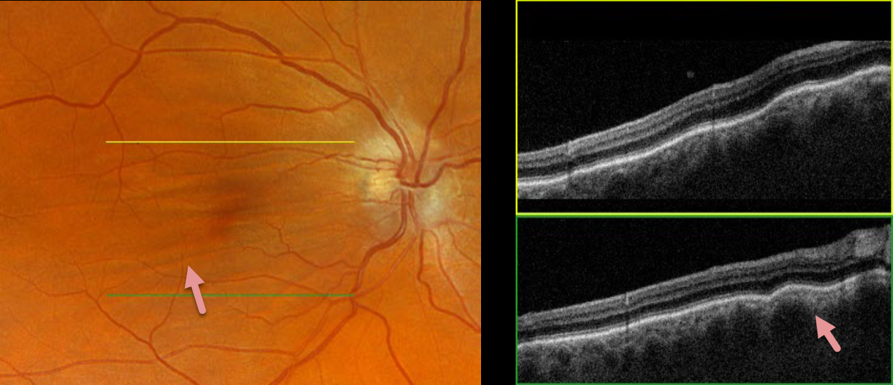
This 52YO male was referred for asymptomatic findings on retinal examination in his right eye. There was a history of refractive amblyopia OD from being a +3-diopter hyperope. He was emmetropic in his normal left eye.
Optos color RGB imaging shows horizontal chorioretinal folds throughout the right macula. These folds are prominently seen on Triton swept-source OCT. Orbital MRI was unremarkable.
Learning Points:
Chorioretinal folds appear clinically as often subtle horizontal and oblique subretinal folds. There are extensive intra- and extra-ocular causes for chorioretinal folds. The typical findings in idiopathic chorioretinal folds are a mild/moderate hyperopic prescription and bilateral, symmetric folds. The folds are thought to result from flattening of the macular sclera.
When unilateral, an MRI is often indicated to rule out an orbital mass. Typical idiopathic chorioretinal folds require no workup and usually cause no symptoms.



