

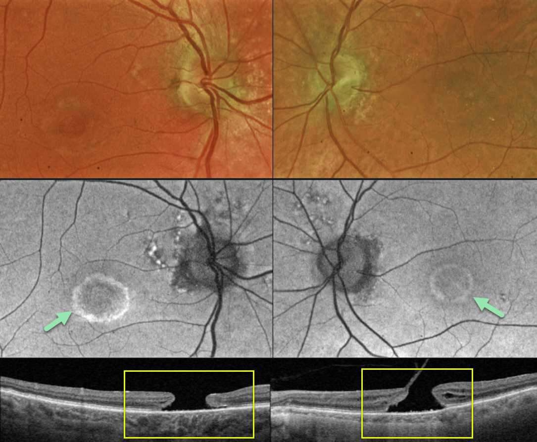
This 83YO male presented with a history of chronic bilateral vision loss. Vision was 20/150 OD and 20/200 OS.
Optos color RG imaging shows a bull’s eye pattern of bilateral pigmentary changes along with a large macular hole OU. Fundus autofluorescence (FAF) shows the bull’s-eye lesions as hyper-FAF. Triton swept-source OCT confirms bilateral macular holes.
Our patient has bilateral chronic macular holes with secondary RPE depigmentation, resulting in a bull’s-eye appearance. Due to the chronicity of the holes, observation was recommended.
Learning Points:
Bull’s eye maculopathy is characterized by a rim of subretinal pigment loss and outer retinal atrophy, most commonly found in inherited retinal diseases (usually ABCA4 disorders) and hydroxychloroquine/chloroquine toxicity.



