

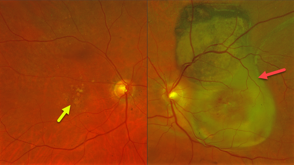
This 78YO female presented with at least several months of vision loss in her left eye. Vision was 20/40 OD and light perception OS. She was examined 2 years earlier with 20/400 vision OS from macular atrophy.
Optos color RG imaging OS shows a variably pigmented and slightly thickened subretinal lesion extending from the superior macula into the superior midperiphery. Subretinal fibrosis extends through the macula, along with retinal blood dots. Intermediate AMD with mixed drusen was noted OD.
Learning Points:
Suprachoroidal, subretinal, or sub-RPE blood can rarely simulate a uveal malignant melanoma. We suggested a trial of monthly anti-VEGF therapy for this treatment naïve neovascular AMD lesion, although the visual prognosis remains quite guarded given the extent of submacular fibrosis.



