

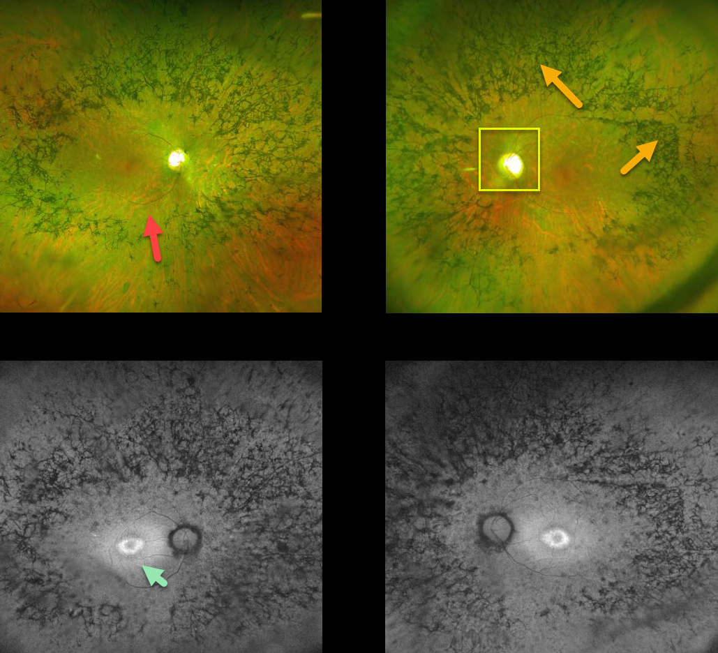
This patient with retinitis pigmentosa (RP)surprisingly has good central vision 20/30 OU.
The fundus photos show all the classic findings for RP, including attenuated retinal vessels, bone spicules (intraretinal migration of the RPE along retinal capillaries), and optic disc pallor.
Fundus autofluorescence for this patient shows a ring of hyperautofluoresence around each central macula. This ring identifies at-risk retina for future degeneration and can help monitor the progression of this disease.
Learning Points:
Retinitis pigmentosa is a retinal degenerative disorder that causes nyctalopia and progressive vision loss.
Photoreceptors normally inhibit RPE cells, so any process with photoreceptor loss can cause similar localized RPE migration.



